Timeline of the development of endoscopic ultrasound (EUS) and transesophageal echocardiography (TEE)
Timeline of the progress in EUS and TEE

Another timeline and references
1971 Side CD and Gosling RG. Transesophageal continuous wave Doppler (ref.1)
1976 Lutz H et al. Transgastric A mode echo (ref.2)
1976 Frazin L et al. Transesophageal M-mode echo (ref.3)
1977 Hisanaga K et al. Transesophageal real-time 2-D sector scanner using a flexible tube (ref.4, ref.5, ref.9, ref.11, ref.12, ref.13) Examination of human. Term "Transesophageal" was first used in this paper. On the other hand, Frazin used Term "esophageal".
1978 Hisanaga K et al. EUS. (Transgastric real-time sector scanner with a gastrofiberscope) (ref.7, ref.8, ref.14) Examination of human, optic guidance.
1978 Hisanaga K et al. Transesophageal 2-D linear scanner. (ref.6, ref.29)
1978 Hisanaga K et al. Transesophageal pulsed Doppler with M-mode. (ref.10, ref.19, ref.39)
1979 Hisanaga K et al. EUS, Transgastric high speed rotating scanner using a flexible tube. (ref.16, ref.18, ref.24, ref.25, ref.27) Human examination. Image of pancreas. (ref.18)
1979 Hisanaga K et al. Transesophageal high speed rotating scanner using a flexible tube. (ref.15, ref.17, ref.18, ref.23, ref.26, ref.28, ref.36, ref.37, ref.38) Human examination.
1979 Hisanaga K et al. Transesophageal high speed rotating scanner for oblique scan. (ref.13, ref.22, ref.34) Four chamber view including apex.
1980 Hisanaga K et al. Filling stomach with water method in EUS examination. (ref.30, ref.31, ref.33) Optic guidance, very important report
1980 Dimagno EP et al. Linear EUS. (animal, dog) (ref.20) Not human but animal
1980 Strohm WD et al. EUS, transgastric mechanical scanner with a fiberscope by Olympus. (ref.32) Examination of human. Optic guidance. Rotating mirror system.
1981 Aibe T et al. EUS (Rotating mirror system with a fiberscope by Olympus) (ref.22)
Optic guidance. Human examination.
1981 Yamanaka J et al. Linear array EUS with a fiber endoscope by Toshiba. Human
examination. Optic guidance. (ref.37A, ref.42) One year before Dimagno.(ref.43).
1982 Schiller NB et al. Transesophageal phased array sector scanner. (ref.39A)
1982 Seward JB et al. Transesophageal phased array sector scanner. (ref.39B) Animal. Dog.
1982 Souquet J et al. Transesophageal phased array sector scanner. (ref.40, ref.41)
Examination of human.
1982 Hanrath P et al. Transesophageal phased array sector scanner. (ref.40A)
1982 Schluter M et al. Transesophageal phased array sector scanner. (ref.40B)
1982 Cahalan MK et al. Transesophageal phased array sector scanner. (ref.40C)
1982 Kremer P et al. Transesophageal phased array sector scanner. (ref.40D)
1982 Aibe T et al. EUS, transgastric rotating transducer scanner with a fiberscope by Olympus. Without rotating mirror. (ref.42A) Human. Optic guidance.
1982 Dimagno EP et al. Linear EUS (human) (ref.43) One year after Yamanaka (ref.42)
1992 Vilman P et al. Transgastric wall fine needle aspiration biopsy in pancreatic disease. (ref.43A)
1993 Roelandt J et al. Transesophageal three-dimensional echo. (ref.44)
1995 Kallimanis G et al. Three-dimensional endoscopic ultrasonography. (ref.44A)
2007 Salgo IS et al. Transesophageal real-time 3D echo. Matrix array probe. (ref.45)
2008 Sugeng L et al. Transesophageal real-time 3D echo. Matrix array probe. (ref.46)
Anybody can reproduce photographies from this website without permission of Hisanaga.
E-mail correspondence [email protected]
References
1. Side CD and Gosling RG Nonsurgical assessment of cardiac function. Nature 232:335-
336, 1971 Transesophageal continuous wave Doppler. Only Doppler signal of Aorta.

From ref.1 Special analysis of Doppler shift signals from descending thoracic Aorta.
Comment : Subject of this investigation was not written in this paper. We could not know whether this image was human's or animal's.
2. Lutz H, Rosch W. Transgastroscopic ultrasonography. Endoscopy 8(4):203-205, 1976 Transgastric A mode only. A-mode is not used now.
3. Frazin L, Tarano JV, Stephanides L, Loeb HS, Kopel L, Gurnar RM. Esophageal echocardiography. Circulation 54:102-108, 1976 Transesophageal M-mode only.
M-mode is time motion display of the Ultrasound wave along a chosen ultrasound line.

From ref.3 Photography of the esophageal transducer. M-mode only.

From ref.3 Panel A shows the external echo of a patient with documented mitral stenosis. Panel B shows the esophageal echo counterpart with reversed orientation.
4. Hisanaga K, Hisanaga A, Nagata K, Yoshida S. A new transesophageal real time two-
dimensional echocardiographic system using a flexible tube and its clinical application. Proceedings of the Japan Society of Ultrasonic in Medicine 32:43-44, 1977
The term "Transesophageal" was first used in this paper. On the other hand Frazin used term "esophageal".

From ref.4 Transesophageal horizontal scan in a normal female.
Flexible tube was used and examinations of human were done.

Kohzoh Hisanaga, MD. (Hisanaga K.) From "People who developed medical ultrasound". p35, Itoh K, Ed., Supplement of Proceedings of 60th Meeting of JSUM 1992. Kohzoh Hisanaga is not only an electronic engineer but also a medical doctor.
Dr. Hisanaga received the Honor Award of the Japanese college of cardiology in 1991 because Dr. Hisanaga developed Transesophageal two-dimensional echocardiography for the first time in history.
5. Hisanaga K, Hisanaga A. A new real-time sector scanning system of ultra-wide angle and real-time recording of entire adult cardiac images --Transesophagus and Trans-chest- wall methods--. In: White DN, Lyons AE (eds.) Ultrasound in Medicine Vol.4. New York, Plenum Press pp391-402, 1978

From ref.5 Insertions of transducer to esophagus and transesophageal ultrasound examinations were performed with patients in left lateral position(left). Typical horizontal scan in a 26-year-old normal adult by using transesophageal method(right).

From ref.5 Transthoracic image. A long axis scan in a 31-year-old normal man. Entire heart image is seen. The endocardium of the left ventricle and the right ventricular anterior wall are seen.

From ref.5 Transesophageal M-mode echograms. These images were recorded in order to identify echo sources of transesophageal cross-sectional images.
6. Hisanaga K, Hisanaga A, Nagata K, Ichie Y, Yoshida S. A new transesophagel high speed linear scanner and its clinical application. Proceedings of the Japan Society of Ultrasonics in Medicine 33:47-48, 1978

From ref.6. Transesophageal vertical scan through mitral valve in a normal adult.
7. Hisanaga K, Hisanaga A. A new trans-digestive-tract scanner with a gastrofiberscope. Proceedings of the 23rd Annual Meeting of American Institute of Ultrasonics in Medicine p.108, November, San Diego, 1978 Human examination. Optic guidance.
This system with a gastrofiberscope was same to that of ref.31. Please see ref.31.
8. Hisanaga K, Hisanaga A. A new trans-digestive-tract scanner with a gastrofiberscope. Reflections 4, 221, 1978 Human examination. Optic guidance.
9. Hisanaga K, Hisanaga A. A new transesophageal high speed sector scanner. Abstracts of 3rd European Congress on Ultrasonics in Medicine (3rd Congress of EFSUME) pp201-205, October 1-5, Bologna 1978

From ref.9 Transesophageal horizontal scan at the level of aortic valve in a normal adult.
10. Hisanaga K, Hisanaga A, Nagata K, Ichie Y, Hibi N, Fukui Y, Nishimura K, Kambe T.
Transesophageal pulsed Doppler echocardiographic system and initial clinical results.
Proceedings of the Japan Society of Ultrasonics in Medicine 34:9-10, 1978
Transesophageal pulsed Doppler with M-mode. Human examination.
11. Hisanaga K, Hisanaga A, Nagata K, Ichie Y, Yoshida S. Transesophageal cross-
Sectional echocardiography -New method for cardiac diagnosis-. (abstr) Jp Circulation J (Circulation Journal) 42:773, 1978
12. Hisanaga K and Hisanaga A. A transesophageal real-time sector scanner with an oil filled cell. Proceedings of the 23rd Annual meeting of American institute of Ultrasound in Medicine, p47, San Diego, 1978

From ref.12 Diagrammatic illustration of the scanner.

From ref.12 Horizontal scan in a normal adult subject.
13. Hisanaga K, Hisanaga A, Ichie Y. A transesophageal ultrasound sector scanner for oblique scan. (abstr) Circulation 60(suppl. II): II-245,1979 Examination of human.
Four chamber view including apex.
14. Hisanaga K, Hisanaga A, Nagata K, Ichie Y. A trans-stomach wall sector scanner
with a gastrofiberscope. Abstracts of 2nd WFUMB, p383, July 22-27, Miyazaki, 1979
Examination of human. Optic guidance.
This system with a gastrofiberscope was same to that of ref.31. Please see ref.31.
15. Hisanaga K, Hisanaga A. A new transesophageal radial scanner using a rotating
flexible shaft and initial clinical results. Proceedings of the 24th Annual Meeting of American Institute of Ultrasound in Medicine p.122, August 27-31, Montreal, 1979

From ref.15 Diagram of transesophageal radial scanner.

From ref.15 A horizontal scan in a normal adult woman by using the transesophageal radial scanner.
From ref.15 A horizontal scan in a normal adult woman by using the transesophageal radial scanner.
16. Hisanaga K, Hisanaga A, Nagata K, Ichie Y. A trans- stomach wall high speed rotating scanner and initial clinical results. Proceedings of the Japan Society of Ultrasonics in Medicine 35:115-116, 1979

Photography of the original paper

From ref.16 Trans-stomach-wall high speed rotating scanner.

From ref.16 Horizontal scan through the left kidney in a normal adult by using the trans-stomach-wall rotating scanner. When near gain is standard. Pancreas is seen as echo free space near the esophagus.
17. Hisanaga K, Hisanaga A, Nagata K, Ichie Y. Cardiac imaging using a transesophageal
ultrasound high speed rotating scanner. Proceedings of the Japan Society of Ultrasonics in Medicine 35:157-158, 1979

Photography of the original paper

From ref.17 Transesophageal high speed rotating scanner.

From ref.17 Transducer and commutator in oil bag.

From ref.17 Horizontal scan at the level of the mitral valve in a patient with severe mitral stenosis by using the transesophageal high speed rotating scanner. Anterior and posterior mitral leaflets are thickened.
From ref.17 Horizontal scan at the level of the mitral valve in a patient with severe mitral stenosis by using the transesophageal high speed rotating scanner. Anterior and posterior mitral leaflets are thickened.
18. Hisanaga K, Hisanaga A. High speed rotating and sector scanners for transesophageal echocardiography and transgastoric sonography. Eizou Jouhou Medical 11:1094-1099, 1979 (in Japanese) Image of pancreas.

From ref.18 Fig.4 Transgastric high speed rotating scanner with a flexible tube.

From ref.18 Fig.11 Horizontal scan at the level of kidney in a normal adult by transgastric high speed rotating scanner. Pancreas is seen very clearly.
19. Hisanaga K, Hisanaga A, Ichie Y, Nishimura K, Hibi N, Fukui Y, Kambe T.
Transesophageal pulsed Doppler echocardiography. Lancet 1:53-54, 1979
Transesophageal pulsed Doppler echo with M-mode
20. Dimagno EP, Regan PT, Wilson DA, Buxton JL, Hattery RR, Suarez JR, Green PS
Ultrasonic endoscope. Lancet 1:629-631, 1980 Note : not human but animal(dog)
Transesophageal electronic linear scanner with a fiberscope.
21. Hisanaga K, Hisanaga A, Nagata K, IchieY, Kambe T. Trans-stomach-wall cross-sectional echhocardiography --examination technique and identification of abdominal cross-sectional image. Proceedings of the Japan Society of Ultrasonics in Medicine 36:
131-132, 1980 Filling stomach with water method during EUS examination.
22. Hisanaga K, Hisanaga A, Nagata K, Ichie Y. A transesophageal high speed rotating scanner for oblique scan and long axis cardiac images including apex. Proceedings of the Japan Society of Ultrasonics in Medicine 36:395-396,1980 Four chamber view including apex.
From ref.22 Inferior oblique scan through the apex in a normal adult. Cross-section is angled downward about 40 degrees from the horizontal plane. The entire heart including the apex is seen.
23. Hisanaga K, Hisanaga A, Kambe T. Detection of atrial septal defect by transesophageal two-dimensional echocardiography. (abstr) Circulation 62(Suppl. III): III-34, 1980
24. Hisanaga K, Hisanaga A. Pancreatic echography using a trans-stomach wall ultrasound rotating scanner. (abstr) Gastroenterology 78:1183, 1980
Examination of human
25. Hisanaga K, Hisanaga A, Kambe T. An endoscopic ultrasound scanner for abdominal echography. (abstr) Gastrointestinal Endoscopy 26:68, 1980 Examination of human
26. Hisanaga K, Hisanaga A, Nagata K, Ichie Y, Isaji F. Transesophageal cross-sectional
echocardiographic diagnosis of Lutembacher's syndrome. Proceedings of the Japan Society
of Ultrasonics in Medicine 37:179-180, 1980

From ref.26 Fig. 4 Transesophageal cross-sectional echocardiogram before surgical repair in a patient with Lutembacher's syndrome. Cross-section is horizontal.
ESO = esophagus, D = defect, LA = left atrium, LV = left ventricle, RA = right atrium, RV = right ventricle.

From ref.26 Fig.5 Transesophageal cross-sectional echocardiogram after surgical repair in a patient with Lutembacher's syndrome. Cross-section is horizontal. Interatrial septum is seen continuously. IAS = interatrial septum.
27. Hisanaga K, Hisanaga A, Nagata K, Ichie Y. High speed rotating scanner for
transgastric sonography. Am J Roentgenol 135:627-629, 1980 Flexible tube was used. Image of pancreas.

From ref.27 Upper Fig. Intragastric high speed rotating scanner. Small transducer in stomach is rotated by flexible rotating shaft and motor at 15-50 cycles/sec. Lower Fig. Transducer and commutator in oil bag.


A B Image of pancreas
From ref.27 Horizontal scans through left kidney in a normal adult with intragastric high speed rotating scanner. A: Left kidney and abdominal aorta are seen clearly. If amplitude of near field is relatively low, pancreas is seen as anechoic space near stomach wall. B: With increasing amplitude of near field, pancreas assumes cloudlike shape.
28. Hisanaga K, Hisanaga A, IchieY, Hibi N, Nishimura K, Kambe T. High speed rotating
scanner for transesophageal cross-sectional echocardiography. Am J Cardiol 46:837-842, 1980

From ref.28 Diagrammatic illustration of the transesophageal high speed rotating scanner. A small transducer in the esophagus is rotated through a full 360° through a flexible shaft by a motor at 15 to 50 cycles /s. Although the small transducer is rotated with great speed in the patient's esophagus, no damage results because the transducer is safely enveloped in an oil bag.

From ref.28 Transesophageal high speed rotating scanner.

From ref.28 Transducer and commutator in oil bag. Sound energy is coupled to and from the transducer through the slip-ring commutator because of the full 360° rotation of the transducer.


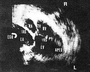
From ref.28 Transesophageal cross-sectional echocardiograms in a patient with mitral stenosis. The cross section is horizontal and shows the heart as viewed from the cardiac apex. A: a frame during diastole and B: a frame during systole. A stenotic mitral orifice (in A) is seen between the tips of the thickened mitral leaflets. The interatrial septum (IAS) is seen without dropout. AML= anterior mitral leaflet, ESO = esophagus, IVS = interventricular septum, LA = left atrium, LV = left ventricle, PML = posterior mitral leaflet, RA = right atrium, RV = right ventricle, TV = tricuspid valve.
29. Hisanaga K, Hisanaga A, Nagata K, Ichie Y. Transesophageal cross-sectional echocardiography. Am Heart J 100:605-609,1980
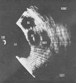
From ref.29 Transesophageal horizontal scan at the level of the aortic valve in a patient with mitral stenosis. The aortic cusps are closed in diastole. Large left atrium is seen. Right ventricular outflow tract is seen anterior to the aorta. AV = aortic valve, RVOT = right ventricular outflow tract.

From ref.29 Transesophageal vertical linear scan through the pulmonary artery in a normal subject. Bifurcation of pulmonary artery and part of ascending aorta are seen.
AO = aorta, PA = pulmonary artery, BI = bifurcation of pulmonary artery.
30. Hisanaga K, Hisanaga A. Endoscopic ultrasound system with optical fiberscope and
examination technique -filing stomach with water method-. Eizou Jouhou Medical Vol.12 No.13, pp773-776, August 1980 (in Japanese) Filling stomach with water method.

From ref.30 Fig.1 Diagrammatic illustration of the transgastric ultrasound sector scanner with a gastrofiberscope.

From ref.30 Fig.3 Transducer in stomach when a normal adult drunk water over 300ml. Comment: There is no dot noise pattern in original photo.

From ref.30 Fig.4 Horizontal scan through the stomach posterior wall in a normal adult by using the transgastric sector scanner with a gastrofiberscope when the normal adult did not drink water.

From ref.30 Fig.5 Horizontal scan through the stomach posterior wall in the same adult by using the transgastric sector scanner with a gastrofiberscope when the adult drunk water over 300ml.
31. Hisanaga K, Hisanaga A, Kambe T. Transgastric sonography and examination technique. Proceedings of the Japan Society of Ultrasonics in Medicine 37:413-414, 1980 Optic guidance. Human examination. Filling stomach with water method.

Photography of the original paper

From ref.31 Fig.2 Transgastiric ultrasound sector scanner with a gastrofiberscope.

From ref.31 Fig.3 Horizontal scan through the stomach posterior wall in a normal adult by using the transgastric sector scanner with gastrofiberscope when the stomach was filled with water. Left kidney is seen clearly. SW = stomach wall, LK = left kidney, V = vertebra, R = right, L = left.
Content of this paper is filling the stomach with water method during EUS examination in order to increase acoustic contact between the stomach wall and transducer. Hisanaga performed this method in 1980.
32. Strohm WD, Phillip J, Hagenmuller F, Classen M. Ultrasonic Tomography by means of ultrasonic fiberendoscope. Endoscopy 12:241-244, 1980 Transgastric mechanical scanner with a fiberscope. Rotating mirror system. Examination of human. Optic guidance.
33. Hisanaga K, Hisanga A. Transgastric endoscopic ultrasonography. Surgical therapy (Geka Chiryo) Vol.44 No.5, 587-593,1981 (in Japanese)
Endoscopic ultrasonography when stomach is filled with water.

From ref.33 Fig.9 Horizontal scan through right and left kidneys by the transgastric scanner when stomach was filled with water over 300ml.
34. Hisanaga K, Hisanaga A. Transesophageal cross-sectional echocardiography with mechanical scanning system. In: Hanrath, Bleifeld and Souquet (eds.) Cardiovascular Diagnosis by Ultrasound, Martinus Nijhoff Publishers pp239-246, 1982

From ref.34 Fig.6 Horizontal scan in a normal adult.
35. Aibe T, Esaki T, Sataka M, Amano H, Kawashima M, Nagatomi Y, Harima K, Azuma M, Maetani N, Ariyama S, Fuji T, Kawamura S, Takemoto T. The investigation of the ultrasonic endoscope. Gastroenterol Endosc 23:728-735, 1981 EUS. (Rotating mirror system with a fiberscope by Olympus) Optic guidance. Human examination.
36. Hisanaga K, Hisanaga A. Measurement of defect size of atrial septal defect by transesophageal two-dimensional echocardiography. Proceedings of the Japan Society of Ultrasonics in Medicine 38:5-6, 1981

Photography of the original paper

From ref. 36 Table 1 Ten patients studied and results.

From ref.36 Comparison of defect size (operative findings) with transesophageal 2D ECHO measurements.

From ref.36 Fig.2 Transesophageal two-dimensional echocardiogram in a patient with an ostium secundum atrial septal defect. Cross-section is horizontal. D = defect.
37. Hisanaga K, Hisanaga A, Isaji F. Transesophageal two-dimensional echocardiographic diagnosis of left atrial myxoma. Proceedings of the Japan Society of Ultrasonics in Medicine 39:457-458, 1981

From ref.37 Transesophageal two-dimensional echocardiogram in a patient with a left atrial myxoma. Cross section is horizontal. A stalk of the tumor is seen clearly. K = stalk

From ref.37 Extracted tumor. Weight of the tumor was 38.5g.
37A. Yamanaka J et al. Proceedings of the Japan Society of Ultrasonics in Medicine 39:409-410, 1981 Electronic linear array EUS with a fiberscope by Toshiba.
Examination of human. Optic guidance.
38. Hisanaga K, Hisanaga A. Findings of coronary artery by transesophageal two-dimensional echocardiography. Proceedings of the Japan Society of Ultrasonics in Medicine 40:171-172, 1982

From ref.38 Transesophageal two-dimensional echocardiogram in a patient with both aortic regurgitation and mitral stenosis. LCA = left coronary artery, RCA = right coronary artery.
39. Hisanaga K, Hisanaga A. Transesophageal pulsed Doppler echocardiography (Third report): Development of system without mirror and initial clinical results. Proceedings of the Japan Society of Ultrasonics in Medicine 40:399-400, 1982 Human.
39A. Schiller NB. Evaluation of cardiac function during surgery by transesophageal 2-dimensional echocardiography. In: Hanrath P, Bleifeld W and Souquet J (eds.) Cardiovascular Diagnosis by Ultrasound, Martinus Nijhoff Publishers pp289-293, 1982
39B. Seward JB, Tajik AJ, Dimagno EP. Esophageal phased-array sector echocardiography. In: Hanrath P, Bleifeld W and Souquet J (eds.) Cardiovascular Diagnosis by Ultrasound, Martinus Nijhoff Publishers pp270-279, 1982 Not human but dog.
40. Souquet J. Phased array transducer technology for transesophageal imaging of the heart-Current status and future aspects-. In: Hanrath P, Bleifeld W and Souquet J (eds).
Cardiovascular Diagnosis by Ultrasound. Martinus Nijhoff Publishers pp251-259, 1982 Notes : human

From ref.40 Mitral valve prolapse.
40A. Hanrath P, Schluter M, Langenstein BA, Polster J, Engel S. Transesophageal horizontal and sagittal imaging of the heart with phased array system. Initial clinical results.
In : Hanrath P, Bleifeld W and Souquet J (eds.) Cardiovascular Diagnosis by ultrasound, Martinus Nijhoff Publishers pp280-288, 1982
40B. Schluter M, Langenstein BA, Polster J, Kremer P, Souquet J, Engel S, Hanrath P. Transesophageal cross-sectional echocardiography with a phased array transducer system. Technique and initial clinical results. Br. Heart J 48:67-72, 1982
40C. Cahalan MK et al. Intraoperative monitoring with two-dimensional echocardiography. Anesthesiology 57: A153, 1982 Transesophageal phased array 2D sector scanner.
40D. Kremer P et al. Intraoperative monitoring of left ventricular performance by transesophageal M-mode and 2-D echocardiography. (abstr) Am J Cardiol 49:956, 1982
41. Souquet J, Hanrath P, Ziteli L, Kremer P, Langestein BA, Schluter M. Transesophageal phased array for imaging the heart. IEEE Transactions on Biomedical Engineering 29:707-712, 1982

Jacques Souquet, PhD. From Eugene A Hessel et. al. Evolution of Perioperative Echocardiography. Anesthesia Key.
42. Yamanaka T, Sakai H, Yoshida Y, Kawamoto C, Ueno N, Kumagai M, Horiguchi M, Nagasawa S, Tanaka M, Seki H, Ido K, Kimura K. Ultrasonic endoscopy for the diagnosis of abdominal lesions. Gastroenterological Endoscopy 24:598-607, April 1982
Electronic linear array endoscopic ultrasonography. Human examination. Optic guidance by fiberscope.
42A. Aibe T, Fuji T, Asagami F, Amano H, Kawashima M, Nagatomi Y, Harima K, Azuma M, Maetani N, Ariyama S, Goto T, Kawaura S, Takemoto T. The investigation of the ultrasonic endoscope (the second report). Gastroenterological endoscopy 24(12):1900-1909, 1982. EUS, transgastric rotating transducer scanner with a fiberscope by Olympus. Without a rotating mirror. Human examination. Optic guidance.
43. Dimagno EP, Regan PT, James EM, Buxton JL. Human endoscopic; ultrasonography.
Gastroenterology 83(4): 824-829, November 1982 Note: Human.
43A. Vilman P, Jacobsen GK, Henriksen FW, Hancke S. Endoscopic ultasonography with guided fine needle aspiration biopsy by pancreatic disease. Gastrointest Endosc 38(2): 172-173, 1992 Transgastric wall fine needle aspiration biopsy.
44. Roelandt J, ten Cate FJ, Bruining N, Salustri A, Vietter WB, Mumm B, van der Putten N. Transesophageal rotoplane echo-CT. A novel approach to dynamic three-dimensional echocardiography. Thoraxcentre J 6:4-8, 1993
44A. Kallimanis G, Garra BS, Tio TL, Krasner B, Al-Kawas FH, Fleischer DE, Zeman RK, Nguyen CC, Benjamin SB. The feasibility of three-dimensional endoscopic ultrasonography: a preliminary report. Gastrointestinal endoscopy 41(3):235-239, 1995
45. Salgo IS. 3D echocardiographic visualization for intracardiac beating heart surgery and intervention. Seminars in Thoracic Cardiovascular Surgery 19:325-329, 2007
Transesophageal real-time 3D echo. Matrix array probe.
46. Sugeng L, Shernan SK, Salgo IS, Weinert L, Shook D, Ramen J, Jeevanandam V, Dupont F, Settlemier S, Savord B, Fox J, Mor-Avi V, Lang RM. Live 3-dimensional transesophageal echocardiography: initial experience using fully-sampled matrix array probe. Journal of the American College of Cardiology 52:446-449, 2008
Transesophageal real-time 3D echo. Matrix array probe.
Apart from gagging, no serious complications were encountered in any of transesophageal and transgastric examinations by Hisanaga.
Medical Doctor Kohzoh Hisanaga himself developed all transesophageal and transgastric systems without the help of any electric companies or organizations except that of Asako Hisanaga.
E-mail address of Hisanaga [email protected]