History of Transesophageal Echocardiography(TEE)
Timeline of the progress in transesophageal echocardiography


When you have time, please see "Timeline of the deveopment of EUS and TEE". Click here
Please see "History of echocardiography". Click here
Please see "History of endoscopic ultrasonography". Click here
Another timeline and references
1971 Side CD and Gosling RG Esophageal continuous wave Doppler echo. (ref.1)
Not examination of the heart but only aorta. Human.
1972 Olson RM et al. Esophageal continuous wave Doppler. (ref.2) Human.
1974 Olson RM et al. Esophageal continuous wave Doppler and pulse echo system. (ref.2A)
1974 Duck FA et al. Monitor during surgery by continuous wave Doppler. Esophageal
continuous wave Doppler. (ref.3) Human.
1975 Tomlin PJ et al. Esophageal continuous wave Doppler. (ref.3A)
1975 Daigle RE et al. Esophageal pulsed Doppler without M-mode. Not a human but dog. (ref.4)
Daigle, coworker of Souquet, visited Dr. Hisanaga and performed human 2D TEE examination in
1979.
1976 Frazin L et al. Esophageal M-mode echo. (ref.5) Not 2D echo but M-mode only.
1977 Matsuzaki M et al. Esophageal M-mode echo. (ref.5A)
1977 Hisanaga K et al. Transesophageal real-time two-dimensional sector scanner using a flexible
tube. (ref.6, ref.7, ref.9, ref.10, ref.11, ref.16, ref.19, ref.22) This is the first transesophageal 2D
scanner in history. Examination of human. The Term "Transesophageal" was first used in this
paper. On the other hand, Frazin used Term "esophageal".
1978 Hisanaga K et al. Transesophageal real-time two-dimensional linear scanner using a flexible
tube. (ref.12, ref.22) Examination of human.
1978 Hisanaga K et al. Transesophageal pulsed Doppler echo with M-mode. (ref.13, ref.13C)
Examination of human. This is the first human transesophageal pulsed Doppler in history.
1978 Hisanaga K et al. EUS (Transgastric sector scanner with a gastrofiberscope). (ref.13A, ref.13B)
Optic guidance. Examination of human.
1979 Hisanaga K et al. Transgastric high speed rotating scanner using a flexible tube.
(ref.13D, ref.13E) Examination of human.
1979 Hisanaga K et al. Transesophageal high speed rotating scanner. (ref.16, ref.17, ref.18, ref.19,
ref.21, ref.24, ref.27, ref.28, ref.39) Examination of human.
1979 Hisanaga K et al. Transesophageal high speed rotating scanner for oblique scan. (ref.14, ref.15,
ref.34) Four chamber view including apex was observed easily by this system.
1979-1981 Hisanaga K et al. Heart images of many kinds of diseases by transesophageal 2D
echocardiography. Atrial septal defect (ref.23, ref.27, ref.34) Lutembacher syndrome(ref.24),
left atrial myxoma(ref.28) and coronary artery (ref.39, ref.40)
1979 Histand MB et al. Transesophageal pulsed Doppler. (ref.20)
1979 Wells MK et al. Transesophageal pulsed Doppler. (ref.20A)
1980 Matsumoto M et al. Intraoperative monitoring by transesophageal M-mode echocardiography.
(ref.25) Not 2D but M-mode.
1981 Schluter M et al. Transesophageal pulsed Doppler with M-mode. (ref.26, ref.32, ref.38C)
1981 Hisanaga K et al. Transesophageal 2-D echo of left atrial myxoma. (ref.28)
1981 Kremer P et al. Transesophageal M-mode echo. (ref.27A) Human.
1981 Hanrath P et al. Transesophageal M-mode echo. (ref.26A, ref.28A)
1982 Hisanaga K et al. Transesophageal 2D echo of coronary artery (ref.39, ref.40)
1982 Schiller NB et al. Transesophageal phased array sector scanner. (ref.29) Monitor in cardiac
surgery. Intraoperative transesophageal 2D echo.
1982 Seward JB et al. Transesophageal phased array sector scanner. (ref.30) Not human but
animal.
1982 Hanrath P et al. Transesophageal phased array sector scanner. (ref.31) Biplane Transesophageal
2D echo
1982 Souquet J et al. Transesophageal phased array sector scanner. (ref.33, ref.36)
1982 Schluter M et al. Transesophageal phased array sector scanner. (ref.35)
1982 Cahalan MK et al. Transesophageal phased array sector scanner. (ref.37)
Intraoperative transesophageal 2D echo.
1982 Roizen MF et al. Transesophageal phased array sector scanner. (ref.37A)
1982 Kremer P et al. Transesophageal phased array sector scanner. (ref.38)
Intraoperative transesophageal 2D echo.
1982 Langenstein BA et al. Transesophageal phased array sector scanner. Transesophageal contrast
2D echo. (ref.38A)
1982 Langenstein BA et al. Transesophageal pulsed Doppler. (ref.38B) Detection of MI and AI by
transesophageal Doppler.
1982 Schluter M et al. Transesophageal pulsed Doppler of MR. (ref.38C)
1982 Reifart N et al. Detection of ASD by mechanical 2D TEE. (Olympus)(ref.39A)
1983 Furuya H et al. Detection of air embolism by M-mode TEE. (ref. 40A)
1983 Thier W et al. 2D-TEE left atrial myxoma. (ref.40AA)
1983 Martin RW et al. Transesophageal Doppler of Air embolism. (ref.40D) Not human but dog.
1983 Ezekowitz MD et al. Transesophageal 2-D echo of left atrial myxoma. (ref.40B)
1984 Bertini A et al. Transesophageal mechanical rotating scanner. (ref.40C)
1984 Curling PE et al. Transesophageal bidirectional phased array echo with temperature
monitoring. (ref.42)
1984 Beaupre PN et al. Intraoperative transesophageal 2D echo. (ref.41A)
1984 Topol EJ et al. Intraoperative transesophageal 2D echocardiography. (ref.43)
Intraoperative transesophageal 2D echo was reported in 1982 already. (ref.29, ref.37, ref.38))
1984 Cucchiara RF et al. Detection of air embolism by transesophageal 2D
echocardiography. (ref.45)
1985 Smith JS et al. Intraoperative detection of myocardial ischemia by TEE. (ref.41)
1985 Topol EJ et al. Intraoperative microbubble detection by transesophageal 2D
echocardiography. (ref.46)
1986 Achenberg W et al. Detection of left atrial appendage thrombus by transesophageal 2D
echocardiography. (ref.47)
1986 Goldman ME et al. Intraoperative transesophageal Doppler flow imaging. Color Doppler. (ref.44)
1986 Takamoto A et al. Transesophageal Doppler flow imaging. Color Doppler. (ref.48)
1987 de Bruijn et al. Intraoperative transesophageal Doppler flow mapping. Color Doppler. (ref.49)
1988 Seward JB et al. Transesophageal 2D echo. Review of TEE. (ref.49A)
1989 Wollschlager et al. Transesophageal dynamic three-dimensional echo(ref.50)
1990 Seward JB et al. Biplane transesophageal 2D echo. (ref.50A) Biplane transesophageal 2D echo
was reported in 1982
1990 Oh JK et al. Management of critical ill patients by TEE. (ref.50B)
1990 Bonsal RC et al. Biplane transesophageal 2D echo. (ref.51) Biplane echo was reported by
Hanrath (ref.31) in 1982.
1990 Omoto R et al. Biplane transesophageal 2D echo. (ref.52) Biplane echo was reported by
Hanrath (ref.31) in 1982.
1992 Roelandt J et al. Multiplane transesophageal 2D echocardiography. (ref.53)
1993 Seward JB et al. Multiplane transesophageal 2D echocardiography. (ref.54)
1993 Roelandt et al. Transesophageal three-dimensional echo (ref.55)
2002 Trambaiolo P et al. Transesophageal tissue Doppler echo. (ref.56)
2003 Cheng MM et al. Transesophageal tissue Doppler echo. (ref.57)
2003 Vitarelli A et al. Transesophageal tissue Doppler echo. (ref.57A)
2007 Salgo IS et al. Transesophageal real-time 3D echo. Matrix array probe. (ref.58)
2008 Desmet M et al. Transesophageal speckle tracking. (ref.58A)
2008 Sugeng L et al. Transesophageal real-time 3D echo. Matrix array probe (ref.59)
2011 Marcucci CE et al. Transesophageal speckle tracking. (ref.59A)
2013 J Am Soc of Echocardiography. Guidelines for performing comprehensive
transesophageal echocardiography. (ref.60)
2022 Fraser AG et al. The most detailed history of echocardiography. (ref.61)
Anybody can reproduce photographies from this website without permission of Hisanaga.
E-mail correspondence [email protected]
Reference
1. Side CD, Gosling RG. Non-surgical assessment of cardiac function. Nature 232:335-
336, 1971 Esophageal continuous wave Doppler echo

From ref.1 Intraesophageal method. Continuous Doppler only. Special analysis of Doppler shift signals from descending thoracic Aorta.
Comment: Subject of this investigation was not written in this paper. We could not know whether this image was human's or animal's.
2. Olson RM, Shelton DK. A nondestructive technique to measure wall displacement in the thoracic aorta. J Appl Physiol 32(1):147-151, 1972. Esophageal continuous wave Doppler echo.
2A. Olson RM, Cooke JP. A nondestructive ultrasonic technique to measure diameter
and blood flow in arteries. IEEE Transactions on Biomedical Engineering 21:168-171, March 1974 Transesophageal continuous wave Doppler and pulse echo system. Human.
3. Duck FA, Hodson CJ, Tomlin PJ. An esophageal Doppler probe for aortic flow
velocity monitoring. Ultrasound in Medicine and Biology 1:233-241,1974
Transesophageal continuous wave Doppler echo. Human.
3A. Tomlin PJ, Duck FA. Transesophageal aortic velography in man. Can Anaesth Soc J 22(5):561-571, 1975 Esophageal continuous wave Doppler.
4. Daigle RE, Miller CW, Histand MB, Macleod FD, Hokanson D. Nontraumatic aortic blood flow sensing by use of an ultrasonic esophageal probe. J Appl Physiol 38:1153-1160, 1975 Examination of dog. Transesophageal pulsed Doppler. Not human but Dog.
Daigle, coworker of Souquet, visited Dr. Hisanaga in Nagoya Japan and performed human 2D TEE examination of normal adult in 1979.
5. Frazin L, Talano JV, Stephanides L, Loeb HS, Kopel L, Gunnar RM. Esophageal
echocardiography. Circulation 54 :102-108,1976 Note: M-mode

From ref.5 Photography of the esophageal transducer.

From ref.5 Panel A shows the external echo of a patient with documented mitral stenosis. Panel B shows the esophageal echo counterpart with reversed orientation.

Leon J. Frazin M.D. From University of Illinois Hospital and Health Sciences System UI Health.
5A. Matsuzaki M et al. Clinical application of esophageal echocardiography to the mitral valve prolapse syndrome. Proceedings of the Japan Society of Ultrasonics in Medicine 31:89-90, 1977 Transesophageal M-mode echo.
6. Hisanaga K, Hisanaga A, Nagata K, Yoshida S. A new transesophageal real time
two-dimensional echocardiographic system using a flexible tube and its clinical application. Proceedings of the Japan Society of Ultrasonics in Medicine 32:43-44, 1977
This is the first report of transesophageal 2D echocardiography in history.
Transesophageal 2D and M-mode echo.

From ref.6 Transesophageal horizontal scan in a 26 years old female.

Kohzoh Hisanaga, MD (Hisanaga K.) From "People who developed medical ultrasound". p35, Itoh K, Ed., Supplement of Proceedings of 60th Meeting of JSUM 1992. Kohzoh Hisanaga is not only an electronic engineer but also medical doctor.
Dr. Hisanaga received the Honor Award of the Japanese college of cardiology in 1991 because Dr. Hisanaga developed Transesophageal two-dimensional echocardiography and endoscopic ultrasound for the first time in history.

The Honor Award of the Japanese college of cardiology
7. Hisanaga K, Hisanaga A. A new real-time sector scanning system of ultra-wide angle
and real-time recording of entire adult cardiac images - Transesophagus and Trans-chest- wall methods -. In: White DN, Lyons AE, eds. Ultrasound in Medicine Vol.4, New York, Plenum Press pp391-402, 1978

From ref.7 Insertions of transducer to esophagus and transesophageal ultrasound examinations were performed with patients in left lateral position. (left)
Typical horizontal scan in a normal adult by using transesophageal method. (right).

From ref.7 Trans-thoracic method. A long axis scan in a 31-year-old normal man. Entire heart image is seen. The endocardiogram of the left ventricle and the right ventricular anterior wall are seen.

From ref.7 Transesophageal M-mode echograms. These images were recorded in order to identify echo sources of transesophageal cross-sectional images shown in Fig.10. Arrows A, B and C of Fig.10A and Fig.10D correspond to Fig.9A, 9B and 9C in each and show directions of M-mode echograms.
8. Hisanaga K, Hisanaga A, Nagata K, Ichie Y, Yoshida S. A new auto-sweeping system for transesophageal M-mode scan and its clinical application. Proceedings of the Japan Society of Ultrasonics in Medicine 33:45-46, 1978

From ref.8 Fig.6 Auto sweeping transesophageal M-mode scan.
9. Hisanaga K, Hisanaga A. A new transesophageal high speed sector scanner.
Abstracts of 3rd European Congress on Ultrasonics in Medicine (3rd Congress of EFSUMB) pp201-205, Bologna, October 1-5, 1978

From ref.9 Transesophageal horizontal scan at the level of aortic valve in a normal adult.
10. Hisanaga K, Hisanaga A, Nagata K, Ichie Y, Yoshida S. Transesophageal cross-sectional echocardiography - New method for cardiac diagnosis -(abstr) Jp Circulation J (Circulation Journal) 42:773, 1978
11. Hisanaga K and Hisanaga A. A transesophageal real-time sector scanner with an oil filled cell. Proceedings of the 23rd Annual Meeting of American Institute of Ultrasound in Medicine, p47, San Diego, 1978

From ref.11 Diagrammatic illustration of the scanner.

From ref.11 Horizontal scan in a normal adult subject.
12. Hisanaga K, Hisanaga A, Nagata K, Ichie Y, Yoshida S. A new transesophageal high speed linear scanner and its clinical application. Proceedings of the Japan Society of Ultrasonics in Medicine 33:47-48. 1978

From ref.12 Transesophageal vertical scan through mitral valve in a normal adult.
13. Hisanaga K, Hisanaga A. A transesophageal pulsed Doppler echocardiographic system and initial clinical results. Proceedings of the Japan Society of Ultrasonics in Medicine 34:9-10, 1978 Transesophageal pulsed Doppler with M-mode. This is the first human transesophageal pulsed Doppler examination in history. Improvement of A.T.L. system.

From ref.13 Mirror and transducer.

From ref.13 Flow pattern in RVOT. Normal adult.
13A. Hisanaga K, Hisanaga A. A new trans-digestive-tract scanner with a gastro-fiberscope. Proceedings of the 23rd Annual Meeting of American Institute of Ultrasound in Medicine p.108, San Diego, November 1978
Examination of human. Trans-stomach wall image of human.
13B. Hisanaga K, Hisanaga A, Nagata K, Ichie Y. A trans-stomach wall sector scanner with a gastrofiberscope. Abstract of 2nd WFUMB p383, Miyazaki, July 22-27, 1979
13C. Hisanaga K, Hisanaga A, Ichie Y, Nishimura K, Hibi N, Fukui Y, Kambe T.
Transesophageal pulsed Doppler echocardiography. Lancet 1:53-54, 1979
Transesophageal pulsed Doppler with M-mode. Improvement of A.T.L. system.
13D. Hisanaga K, Hisanaga A, Nagata K, Ichie Y. A trans- stomach wall high speed rotating scanner and initial clinical results. Proceedings of the Japan Society of Ultrasoics in Medicine 35:115-116, 1979 A flexible tube was used. Examination of human.
13E. Hisanaga K, Hisanaga A, Nagata K, Ichie Y. High speed rotating scanner for
transgastric sonography. Am J Roentgenol 135:627-629, 1980 Examination of human.
14. Hisanaga K, Hisanaga A, Nagata K, Ichie Y. A transesophageal high speed rotating scanner for oblique scan and long axis cardiac images including apex. Proceedings of the Japan Society of Ultrasonics in Medicine 36:395-396, 1980
Four chamber view including apex could be observed easily by this system.

From ref.14 Inferior oblique scan through the apex in a normal adult. Cross-section is angled downward about 40 degrees from the horizontal plane. The entire heart including the apex is seen.
15. Hisanaga K, Hisanaga A, Ichie Y. A transesophageal ultrasound sector scanner for oblique scan (abstr). Circulation 60(suppl. II): II-245,1979
16. Hisanaga K, Hisanaga A. High speed rotating and sector scanners for transesophageal echocardiography and transgastric sonography. Eizou Jouhou Medical 11:1094-1099, 1979 (in Japanese)

From ref.16 Fig 7 Transesophageal horizontal scan in a patient with ASD before surgical repair by using transesophageal sector scanner.
17. Hisanaga K, Hisanaga A. A new transesophageal radial scanner using a rotating flexible shaft and initial clinical results. Proceedings of the 24th Annual Meeting of American Institute of Ultrasound in Medicine p.122, Montreal, August 27-31 1979

From ref.17 Diagram of transesophageal radial scanner.

From ref.17 A horizontal scan in a normal adult woman by using the transesophageal radial scanner.
18. Hisanaga K, Hisanaga A, Nagata K, Ichie Y. Cardiac imaging using a transesophageal ultrasound high speed rotating scanner. Proceedings of the Japan Society of Ultrasonics in Medicine 35:157-158, 1979

Photography of the original paper.

From ref.18 Transesophageal high speed rotating scanner.

From ref.18 Transducer and commutator in oil bag.

From ref.18 Horizontal scan at the level of the mitral valve in a patient with severe mitral stenosis by using the transesophageal high speed rotating scanner. Anterior and posterior mitral leaflets are thickened.
19. Hisanaga K, Hisanaga A, Fukui K, Nagata K, Ichie Y. Atrial septal defect visualization by transesophageal cross-sectional echocardiography. Proceedings of the Japan Society of Ultrasonics in Medicine 35:51-52. 1979

Photography of the original paper

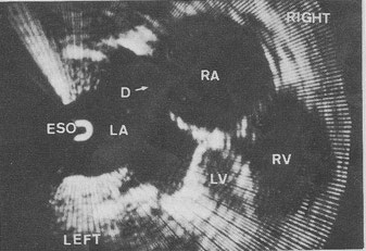
From ref.19 Fig.1 Preoperative horizontal scan at the level of the mitral valve in a patient with atrial septal defect by using a transesophageal high speed sector scanner. (case 1) D = defect

From ref.19 Fig. 3 Postoperative horizontal scan at the level of the mitral valve in the patient as shown in Fig.1 by using a transesophageal high speed rotating scanner. (case 1)
20. Histand MB, Wells MK, Reeves JT, Sodal IE, Adamson HP, Willson JT. Ultrasonic pulsed Doppler transesophageal measurement of aortic hemodynamics in humans. Ultrasonics 17(5):215-218, Sep 1979
20A. Wells MK, Histand MB, Reeves JT, Adamson HP. Ultrasonic transesophageal measurement of hemodynamic parameters in humans. ISA Trans 18(1):57-61, 1979
Transesophageal Doppler echo. Human.
21. Hisanaga K, Hisanaga A, IchieY, Hibi N, Nishimura K, Kambe T. High speed rotating scanner for transesophageal cross-sectional echocardiography. Am J Cardiol 46:837-842, 1980

From ref.21 Diagrammatic illustration of the transesophageal high speed rotating scanner. A small transducer in the esophagus is rotated through a full 360° through a flexible shaft by a motor at 15 to 50 cycles /s. Although the small transducer is rotated with great speed in the patient's esophagus, no damage results because the transducer is safely enveloped in an oil bag.

From ref.21 Transesophageal high speed rotating scanner.

From ref.21 Transducer and commutator in oil bag. Sound energy is coupled to and from the transducer through the slip-ring commutator because of the full 360° rotation of the transducer.


From ref.21 Transesophageal cross-sectional echocardiograms in a patient with mitral stenosis. The cross section is horizontal and shows the heart as viewed from the cardiac apex. A: a frame during diastole and B: a frame during systole. A stenotic mitral orifice (in A) is seen between the tips of the thickened mitral leaflets. The interatrial septum (IAS) is seen without dropout. AML= anterior mitral leaflet, ESO = esophagus, IVS = interventricular septum, LA = left atrium, LV = left ventricle, PML = posterior mitral leaflet, RA = right atrium, RV = right ventricle, TV = tricuspid valve.
22. Hisanaga K, Hisanaga A, Nagata K, IchieY. Transesophageal cross-sectional echocardiography. Am. Heart J 100:605-609, 1980

From ref.22 Transesophageal horizontal scan at the level of the aortic valve in a patient with mitral stenosis. The aortic cusps are closed in diastole. Large left atrium is seen. Right ventricular outflow tract is seen anterior to the aorta. AV = aortic valve, RVOT = right ventricular outflow tract.

From ref.22 Transesophageal vertical linear scan through the pulmonary artery in a normal subject. Bifurcation of pulmonary artery and part of ascending aorta are seen. AO = aorta, PA = pulmonary artery, BI = bifurcation of pulmonary artery.
23. Hisanaga K, Hisanaga A, Kambe T. Detection of atrial septal defect by transesophageal two-dimensional echocardiography. (abstr) Circulation 62(suppl Ⅲ):Ⅲ-34, 1980

From ref.23 Original abstract, Circulation (Suppl Ⅲ)Ⅲ-34,1980
24. Hisanaga K, Hisanaga A, Nagata K, Ichie Y, Isaji F. Transesophageal cross-sectional echocardiographic diagnosis of Lutembacher syndrome. Proceedings of the Japan Society of Ultrasonics in Medicine 37:179-180, 1980
From ref.24 Fig. 4 Transesophageal cross-sectional echocardiogram before surgical repair in a patient with Lutembacher syndrome. Cross-section is horizontal.
ESO = esophagus, D = defect, LA = left atrium, LV = left ventricle, RA = right atrium, RV = right ventricle.

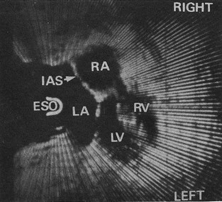
From ref.24 Fig.5 Transesophageal cross-sectional echocardiogram after surgical repair in a patient with Lutembacher's syndrome. Cross-section is horizontal. Interatrial septum is seen continuously. IAS = interatrial septum.
25. Matsumoto M, Oka Y, Strom J, Frishman W, Kadish A, Becker RM, Frater RW, Sonnenblick
EH. Application of transesophageal echocardiography to continuous intraoperative
monitoring of left ventricular performance. Am J Cardiol 46(1):95-105, 1980
Not transesophageal 2D echo but M-mode echo.
26. Schluter M, Langenstein BA, Hanrath P, Kremer P, Bleifeld W. Mitral regurgitation detected by transesophageal pulsed Doppler echocardiography. (abstr) Eur Heart J 2(suppl A) 114, 1981 Transesophageal pulsed Doppler by improvement of A.T.L. system.
26A. Hanrath P, Kremer P, Langenstein BA, Matsumoto M, Bleifeld W. Transosophageale Echokardiographie, Ein neues Verfahren zur dynamischen Ventrikelfunktionsanalyse. Deutsche Med Wschr 156:523, 1981. Transesophageal M-mode echo.
27. Hisanaga K, Hisanaga A, Nagata K, Ichie Y. Measurement of defect size of atrial septal defect by transesophageal two-dimensional echocardiography. Proceedings of the Japan Society of Ultrasonics in Medicine 38:5-6, 1981

Photography of original paper

From ref.27 Table 1 Ten patients studied and results.

From ref.27 Fig.2 Transesophageal two-dimensional echocardiogram in a patient with an ostium secundum atrial septal defect. Cross-section is horizontal. D = defect.
From ref.27 Fig.3 Same patient. Transesophageal two-dimensional echocardiogram after repair of the atrial septal defect. The repaired interatrial septum (IAS) is imaged almost continuously.
27A. Kremer P, Hanrath P, Langenstein BA, Matsumoto M, Tams C, Bleifeld W. The evaluation of left ventricular function at rest and during exercise by transesophageal echocardiography in aortic insufficiency. (abstr) Am J Cardiol 47:412, 1981 Transesophageal M-mode.
28. Hisanaga K, Hisanaga A, Isaji F. Transesophageal two-dimensional echocardiographic diagnosis of left atrial myxoma. Proceedings of the Japan Society of Ultrasonics in Medicine 39:457-458, 1981
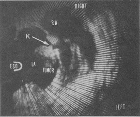
From ref.28 Fig.4 Transesophageal two-dimensional echocardiogram in a patient with a left atrial myxoma. Cross section is horizontal. A stalk of the tumor is seen clearly. K = stalk.

From ref.28 Fig.5 Extracted tumor. Weight of the tumor was 38.5g.
28A. Hanrarh P, Matsumoto M. Transesophageal echocardiography: A new method for the evaluation of left ventricular performance during dynamic exercise. Edited by A Kurjak in: Recent Advances in Ultrasound Diagnosis 3: 393, 1981. Transesophageal M-mode echocardiography.
29. Schiller NB. Evaluation of cardiac function during surgery by transesophageal 2-dimensional echocardiography. In: Hanrath P, Bleifeld W, Souquet J (eds.) Cardiovascular Diagnosis by Ultrasound, Martinus Nijhoff Publishers pp289-293, 1982
Intraoperative transesophageal 2D echocardiography.
30. Seward JB, Tajik AJ, Dimagno EP. Esophageal phased-array sector echocardiography: an anatomic study. In: Hanrath P, Bleifeld W, Souquet J (eds.) Cardiovascular Diagnosis by Ultrasound, Martinus Nijhoff Publishers pp270-279, 1982 Not human but animal.
31. Hanrath P, Schluter M, Langenstein BA, Polster J, Engel S. Transesophageal
horizontal and sagittal imaging of the heart with phased array system. Initial clinical results.
In: Hanrath P, Bleifeld W and Souquet J (eds.) Cardiovascular Diagnosis by ultrasound,
Martinus Nijhoff Publishers pp280-288, 1982
32. Schluter M, Langenstein BA, Hanrath P. Transesophageal pulsed Doppler echocardiography in mitral and aortic regurgitation. In: Hanrath P, Bleifeld W and Souquet J (eds.) Cardiovascular Diagnosis by ultrasound, Martinus Nijhoff Publishers pp132-140, 1982 Transesophageal Pulsed Doppler with M-mode by improvement of A.T.L. system.
33. Souquet J, Hanrath P, Ziteli L, Kremer P, Langenstein BA, Schluter M. Transesophageal phased array for imaging the heart. IEEE Trans Biomed Eng 29:707-712, 1982

Jacques Souquet, PhD. From Eugene A Hessel et. al. Evolution of Perioperative Echocardiography. Anesthesia Key.
34. Hisanaga K, Hisanaga A. Transesophageal cross-sectional echocardiography with a mechanical scanning system. In: Hanrath P, Bleifeld W and Souquet J(eds.) Cardiovascular Diagnosis by Ultrasound, Martinus Nijhoff Publishers pp239-246, 1982

From ref.34 Transesophageal horizontal scan in a normal adult.
35. Schluter M, Langenstein BA, Polster J, Kremer P, Souquet J, Engel S, Hanrath P.
Transesophageal cross-sectional echocardiography with a phased array transducer system.
Technique and initial clinical results. Br Heart J 48:67-72, 1982
36. Souquet J. Phased array transducer technology for transesophageal imaging of the heart -current status and future aspects-. In: Hanrath P, Bleifeld W and Souquet J(eds.) Cardiovascular Diagnosis by Ultrasound, Martinus Nijhoff Publishers pp251-259, May 1982

From ref. 36 Mitral valve prolapse.
RE Daigle, coworker of Souquet, visited Hisanaga's hospital and performed human 2D TEE examination in 1979.
37. Cahalan MK et al. Intraoperative monitoring with two-dimensional echocardiography. Anesthesiology 57: A153, 1982 Transesophageal phased array 2D echocardiography.
Intraoperative monitoring with transesophageal 2D echo.
37A. Roizen MF, Kremer P, Cahalan M, Shiller N, Stoney RJ, Sohn YJ, Cronnelly R, Ehrenfeld WJ. Monitoring with transesophageal echocardiography: Patients undergoing supraceliac aortic occlusion. Anesthesiology 57(3): A154, 1982
38. Kremer P et al. Intraoperative monitoring of left ventricular performance by transesophageal M-mode and 2-D echocardiography. (abstr) Am J Cardiol 49(4):956, 1982
Transesophageal phased array 2D echocardiography.
38A. Langenstein BA, Hanrath P, Polster J, Kremer P, Engel S, Souquet J. Detection of atrial septal defect by transesophageal cross-sectional contrast echocardiography. (abstr) Am J Cardiol 49(4):956, March 1982
38B. Langenstein BA, Schluter M, Hanrath P, Kremer P, Bleifeld W. Detection of mitral and aortic regurgitation by transesophageal pulsed Doppler echocardiography. (abstr.) Am J Cardiol 49:959, 1982
38C. Schluter M, Langenstein BA, Hanrath P, Kremer P, Bleifeld W. Assessment of transesophageal pulsed Doppler echocardiography in the detection of mitral regurgitation.
Circulation 66:784-789, 1982
39. Hisanaga K, Hisanaga A. Findings of coronary artery by transesophageal two-dimensional echocardiography. Proceedings of the Japan Society of Ultrasonics in Medicine 40:171-172, 1982

From ref.39 Transesophageal two-dimensional echocardiogram in a patient with both aortic regurgitation and mitral stenosis. LCA = left coronary artery, RCA = right coronary artery.
39A. Reifart N, Strohm WD. Detection of atrial septal defects by transesophageal two-dimensional echocardiography with a mechanical sector scanner. In: Hanrath P, Bleifeld W and Souquet J (eds.) Cardiovascular Diagnosis by Ultrasound, Martinus Nijhoff Publishers pp247-250, May 1982
40. Hisanaga K, Hisanaga A. A visualization of coronary artery by transesophageal two-dimensional echocardiography (2nd report). Proceedings of the Japan Society of Ultrasonics in Medicine 41:715-716, 1982
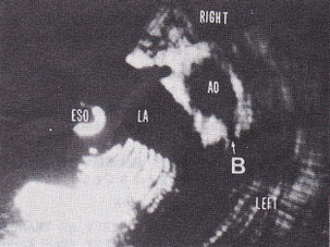
From ref.40 Fig.3 Transesophageal two-dimensional echocardiogram. B=left coronary artery
40A. Furuya H, Suzuki T, Okumura F, Kishi Y, Uefuji T. Detection of Air Embolism by Transesophageal Echocardiography. Anesthesiology 58:124–129, February 1983
40AA. Thier W, Schluter M, Krebber HJ, Polonius MJ, Kloppel G, Becker K, Hanrath P. Cysts in left atrial myxomas identified by transesophageal cross-sectional echocardiography. Am J Cardiol 51(10):1793-1795, June 1983
40B. Ezekowitz MD, Smith EO, Rankin R, Harrison LH Jr. Krous HF. Left atrial mass: diagnostic value of transesophageal 2-dimensional echocardiography and indium-Ⅲ platelet scintigraphy. Am J Cardiol 51(9):1563-1564, 15 May 1983
Detection of left atrial myxoma by 2-D TEE.
40C. Bertini A, Masotti L, Zuppiroli A et al. Rotating probe for trans-oesophageal cross-sectional echocardiography. J Nucl Med Allied Sci 28:115-121, 1984
40D. Martin RW, Colley PS. Evaluation of transesophageal Doppler detection of air embolism in dogs. Anesthesiology 58(2):117-123, February 1983
41. Smith JS, Cahalan MK et al. Intraoperative detection of myocardial ischemia in high- risk patients: electrocardiography versus two-dimensional transesophageal echocardiography. Circulation 72(5): 1015-1021, 1985
41A. Beaupre PN, Kremer PF et al. Intraoperative detection of changes in left ventricular segmental wall motion by transesophageal two-dimensional echocardiography. Am Heart J 107(5): 1021-1023, 1984
42. Curling PE, Newsome LR et al. 2D-transesophageal echocardiography: a bidirectional phased array probe with temperature monitoring. (abstr) Anesthesiology 61(3A):159, 1984 When 2D TEE examination, operator should be aware that probe temperature will reach 41-42℃ with two minutes.
43. Topol EJ, Weiss JL, Guzman PA, Dorsey-Lima S, Blanck TJ, Humphrey LS, Baumgartner WA, Flaherty JT, Reitz BA. Immediate improvement of dysfunctional myocardial segments after coronary revascularization: detection by intraoperative transesophageal echocardiography. J Am Coll Cardiol 4(6): 1123-1134, 1984
Intraoperative transesophageal echo was reported in 1982. (ref.29, ref.37, ref.38)
44. Goldman ME, Thys D, Ritter S, Hittel Z, Kaplan J. Transesophageal real-time Doppler flow imaging: a new method for intraoperative cardiac evaluation (abstract). J Am Coll Cardiol 7:1, 1986 Intraoperative real-time Doppler flow mapping.
45. Cucchiara RF, Nugent M et al. Air embolism in upright neurosurgical patients: detection and localization by two-dimensional transesophageal echocardiography. Anesthesiology 60: 353-355, 1984
46. Topol EJ, Humphrey LS et al. Value of intraoperative left ventricular microbubbles detected by transesophageal two-dimensional echocardiography in predicting neurologic outcome after cardiac operations. Am J Cardiol 56(12): 773-775, 1985
47. Achenberg W, Schluter M, Klemer P et al. Transesophageal two-dimensional echocardiography for the detection of left atrial appendage thrombus. J Am Coll Cardiol 7(1):163-166, 1986
48. Takamoto S, Kyo S, Matsumura M, Hojo H, Yokote Y, Omoto R. Total visualization of thoracic dissecting aortic aneurysm by transesophageal Doppler color flow mapping (abstract). Circulation 74(Suppl II): II–132, 1986
49. de Bruijn NP, Clements FM, Kisslo JA. Intraoperative transesophageal color flow mapping: initial experience. Anesth Analg 66:386–390, 1987
49A. Seward JB, Khandheria BK et al. Transesophageal echocardiography: technique, anatomic correlations, implementation and clinical applications. Mayo Clinic Proceedings 63(7): 649-680, 1988 Review of TEE.
50. Wollschlager H, Zeiher AM, Klein HP, Kasper W, Wollschlager S, Geibel A, Just H.
"Transesophageal echo computer tomography (ECHO-CT): a new method of dynamic 3-D
reconstruction of the heart." (in German) Biomed Tech (Berl) 34 Suppl:10-1, 1989
50A. Seward JB, Khandheria BK et al. Biplane transesophageal echocardiography: anatomic correlations, image orientation and clinical application. Mayo clinic Proceedings 65: 1193-1213, 1990
50B. Oh JK, Seward JB, Khandheria BK, Gersh BJ, McGregor CG, Freeman WK, Sinak LJ, Tajik AJ Transesophageal echocardiography in critically ill patients. Am J Cardiol 66(20):1492-1495, 1990
51. Bansal RC, Shakudo M, Shah PM et al. Biplane transesophageal echocardiography: technique, image orientation, and preliminary experience in 131 patients. Journal of the American Society of Echocardiography 3(5): 348-366, 1990
Biplane echo was reported by Hanrath (ref.28B) in 1982
52. Omoto R, Kyo S, Matsumura M, Adachi H, Masuyama M, Matsunaka T. New direction of biplane transesophageal echocardiography with special emphasis on real-time biplane imaging and matrix phased-array biplane transducer. Echocardiography 7(6):691-698, 1990 Biplane echo was reported by Hanrath (ref.28B) in 1982
53. Roelandt J, Thomson IR, Vletter WB, Brommersma P, Bom N, Linker DT. Multiplane transesophageal echocardiography: latest evolution in an imaging revolution. Journal of the American Society of Echocardiography 5(4):361-367, 1992
Multiplane transesophageal 2D echo.
54. Seward JB, Khandheria BK, Freeman WK, Oh JK, Enriquez-Sarano M, Miller FA, Edwards WD, Tajik AJ. Multiplane transesophageal echocardiography: image orientation, examination technique, anatomic correlations and clinical applications. Mayo clinic Proceedings 68:523-551, 1993
55. Roelandt J, ten Cate FJ, Bruining N, Salustri A, Vietter WB, Mumm B, van der Putten N. Transesophageal rotoplane echo-CT. A novel approach to dynamic three-dimensional echocardiography. Thoraxcentre J 6:4-8, 1993
56. Trambaiolo P, Salustri A et al. Assessment of left atrial appendage wall velocities by transesophageal tissue Doppler echocardiography: a clinical study in patients with sinus rhythm. J Am Soc Echocardiogr 15(5): 425-432, 2002
57. Cheng MM, Li J, White PA et al. Doppler tissue echocardiography: can trans-esophageal echocardiography be used to acquire functional data? J Am Soc Echocardiogr 16(7):732-737, 2003
57A. Vitarelli A, Conde Y, Cimmino E, Colantonio R, D’Angeli I, Stellato S. Transesophageal echocardiography with tissue Doppler imaging in the assessment of global and regional systolic and diastolic right ventricular function in atrial septal defects. Journal of cardiac failure 9:5 Suppl 1, S23, October, 2003
58. Salgo IS. 3D echocardiographic visualization for intracardiac beating heart surgery and intervention. Seminars in Thoracic Cardiovascular Surgery 19:325-329, 2007
Transesophageal real-time 3D echo. Matrix array probe.
58A. Desmet M, Bowry R, Tousignant C. Use of transesophageal speckle tracking echocardiography to evaluate right ventricular longitudinal strain during cardiac surgery. Canadian Journal of Anaesthesia 55(S1):4608071-4608072, 2008
59. Sugeng L, Shernan SK, Salgo IS, Weinert L, Shook D, Ramen J, Jeevanandam V, Dupont F, Settlemier S, Savord B, Fox J, Mor-Avi V, Lang RM. Live 3-dimensional transesophageal echocardiography initial experience using fully-sampled matrix array probe. Journal of the American College of Cardiology 52:446-449, 2008 Transesophageal real-time 3D echo. Matrix array probe.
59A. Marcucci CE, Samad Z, Rivera J, Adams DB, Philips-Bute BG, et al. A comparative evaluation of transesophageal and transthoracic echocardiography for measurement of left ventricular systolic strain using speckle tracking. J Cardiothorac Vasc Anesth. 26(1):17–25, 2011
60. Harn RT, Abraham T et al. Guidelines for performing comprehensive transesophageal echocardiographic examination: recommendations from the American Society of Echocardiography and the Society of Cardiovascular Anesthesiologists. J Am Soc Echocardiogr 26:921-64, 2013
61. Fraser AG, Monaghan MJ, van der Steen AFW, Sutherland GR. A concise history of echocardiography: timeline, pioneers, and landmark publications. European Heart Journal - Cardiovascular Imaging 00:1–14, 2022 The most detailed history of echocardiography.

From ref.61 Page 1. Graphical abstract. Hisanaga’s research performance of transesophageal imaging is written in this abstract.
Apart from gagging, no serious complications were encountered in any
of the transesophageal and transgastric examinations by Hisanaga.
Medical Doctor Hisanaga himself developed all transesophageal and transgastric systems without help of any electric companies or organizations except that of Asako Hisanaga.