History of Echocardiography
Origin of Echocardiography
Development of Echocardiography
History of Cardiac Ultrasound
Timeline of the progress in echocardiography


Another time line and references
1954 Edler I and Hertz Ch. M-mode echo (UCG) (ref.1, ref.3, ref.69)
1956 Satomura S. Continuous wave Doppler echo (ref.2, ref.4, ref.4A)
1957 Wild JJ et al. Visualization of the excised human heart by ultrasound. (ref.2A)
1960 Cieszynski T. Catheter head ultrasound probe. Animal (dog) A-mode. (ref.5)
1963 Olofsson S. Ultrasonic optical mirror system. 2D image of isolated heart of calf. (ref.6)
1963 Hertz CH and Asberg AG 5th International Conference on Medical Electronics in Liege, Belgium in
July 1963 (ref.7, ref.8) The first report of 2D images of the human heart in vivo.
1963 Omoto R et al. Ultrasonic intravenous sonde (ref.9, ref.14)
1963 Joyner CR Jr et al. Assessment of mitral valve by M-mode. (ref.9A)
1964 Hertz CH. First full paper of real-time two-dimensional echocardiography of the living human heart
according to 5th international conference in Belgium (ref.8) ic visualization of soft tissues. (ref.10)
1965 Feigenbaum H et al. Diagnosis of pericardial effusion of heart by M-mode. (ref.11A)
1965 Asberg AG. Ultrasonic images of the living heart (ref.11) Human.
1967 Hertz CH. Ultrasonic images of the living heart (ref.12)
1967 Asberg AG. Ultrasonic cinematography of the living heart (ref.13)
1967 Baker DW. Pulsed Doppler (ref.16, ref.23)
1967 Ebina T et al. Compound electrocardiograph-gated cross-sectional imaging of the heart (ref.15)
1967 Somer JC. Phased array sector scanner (ref.17, ref.18, ref.19)
1968 Joyner C. Contrast echo (ref.20) Contrast echo was found by Joyner for the first time in history.
1968 Gramiak R and shah PM. Contrast echo. (ref.21, ref.21A)
1969 Light LH. Observing blood-flow in human aorta by Doppler. (ref. 18A)
1969 Wells PNT. Range gated ultrasonic Doppler system (ref.22)
1971 Side CD and Gosling RG. Transesophageal continuous wave Doppler echo (ref.24)
1971 Bom N et al. Linear B-mode echo (ref.25, ref.27)
1972 King DL. Compound electrocardiograph-gated cross-sectional image of the heart. (ref.26)
1972 Johnson ML et al. M-mode echo during mitral valve surgery. (ref.26A)
1973 King DL. Real-time linear 2D heart echo. (ref.27A)
1973 Griffith JM and Henry WL. Mechanical sector scanner (ref.28, ref.28A, ref.30)
1973 Johnson SL et al. Pulsed Doppler echo. (ref.28B)
1973 Gramiak R, Waag R, Simon W. Cine ultrasound cardiography (ref.29)
1974 von Ramm OT, Thurstone FL et al. Phased array sector scanner (ref.31, ref.32)
1974 Teichholz LE et al. B-scan ultrasonography of the heart. (ref.32A)
1974 Eggleton RC et al. Mechanical sector scanner (ref.33, ref.34)
1975 Huntsman LL et al. Observing human aortic blood-flow Doppler. (ref.33A)
1975 Holm HH et al. Mechanical 2D scanner. (ref.33B)
1976 Weyman AE et al. Left main coronary image by 2D echo. (ref.34C)
1976 Nealeigh RC et al. A venous pulse Doppler catheter tip flowmeter. (ref.34A)
1976 Holen J et al. Determination of pressure gradient with Doppler echo. (ref.34B)
1976 Frazin L et al. Esophageal M mode echo (ref.35)
1977 Hisanaga K et al. Ultrawide angle high speed sector scanner (ref.35A, ref.37)
Entire adult heart image.
1977 Hisanaga K et al. Transesophageal 2D sector scanner (ref.36, ref.37, ref.38)
This is the first transesophageal 2D scanner in history. The term “Transesophageal” was first used in
this paper. On the other hand, Frazin used term “esophageal”.
1978 Hatle L et al. Assessment of pressure drop in MS by Doppler. (ref.36A)
1978 Hisanaga K et al. Transesophageal 2D linear scanner (ref.39, ref.51)
1978 Brandestini MA. M-mode and Digital multigated Doppler flowmeter (ref.40)
1978 Hisanaga K et al. Transesophageal pulsed Doppler with M-mode (ref.41, ref.42)
1978 Griffith JM et al. Combined cardiac imaging and Doppler flow measurement. (ref.41A)
1978 Barash PG et al. Monitoring left ventricular function by M-mode in anesthetized patient. (ref.41B)
1979 Hisanaga K et al. Transesophageal high speed rotating scanner (ref.43, ref.44, ref.50, ref.52, ref.53,
ref.57)
1979 Hisanaga et al. Transesophageal high speed rotating scanner for oblique scan. (ref.45, ref.48,
ref.57) Four chamber view including apex.
1979 Holen J, Simonsen S Determination of pressure gradient of MS with Doppler echo. (ref.46)
1980 Hatle L et al. Assessment of AS by Doppler. (ref.46A)
1980 Spotnitz HM et al. Intraoperative 2D echo. (ref.51A, ref.52B)
1981 Sahn DJ Intraoperative 2D echo. (ref.52A)
1982 Ghosh A et al. Three-dimensional echo using rotational method (ref.54)
1982 Schiller NB et al. Transesophageal 2D phased array sector scanner. (ref.55)
1982 Seward JB et al. Transesophageal 2D phased array sector scanner. Dog. (ref.55A)
1982 Souquet J et al. Transesophageal 2D phased array sector scanner. (ref.55B, ref.56)
1982 Hanrath P et al. Transesophageal 2D phased array sector scanner. (ref.56A)
1982 Schluter M et al. Transesophageal 2D phased array sector scanner. (ref.56B)
1982 Bommer WJ et al. 2D color flow Doppler. (ref.58)
1982 Namekawa K et al. 2D color flow Doppler with a mechanical scanner. (ref.59, ref.60)
1982 Cahalan MK et al. Transesophageal phased array sector scanner. (ref.56C)
1982 Kremer P et al. Transesophageal phased array sector scanner. (ref.56D)
1982 Langenstein BA et al. Transesophageal phased array sector scanner. (ref.56E)
2D images of ASD
1984 Yock PG et al. Right ventricular systolic pressure by Doppler. (ref.60A)
1985 Kasai C et al. 2D color flow Doppler with a phased array scanner. (ref.61)
1988 Pandian NG et al. Intraluminal ultrasound image of blood vessels. (ref.61AA)
1988 Trahey GE, Smith SW et al. Properties of acoustical speckle. (ref.61A, ref.61B)
1989 Bertrand M, Meunier J et al. Speckle tracking method. (ref.61C, ref.61D)
1989 Wollschlager H et al. Transesophageal dynamic three-dimensional echo. (ref.62)
1991 von Ramm OT et al. Real-time three-dimensional ultrasound (ref.64)
1991 Nissen SE et al. Intracoronary 2D echo (ref.63)
1992 McDicken WN et al. Color Doppler myocardial imaging. (ref.64A) Tissue Doppler Imaging.
1993 Wells PNT. History of echocardiography. (ref.64AA)
1993 Roelandt J et al. Transesophageal three-dimensional echo (ref.66)
1994 Sutherland GT et al. Color Doppler myocardial imaging. (ref.65A)
1995 Tei C et al. Tei index. (ref.64B, ref.64C)
1995 Paika P et al. Tissue Doppler imaging. (ref.65B)
1995 Miyatake K et al. Tissue Doppler imaging. (ref.65C)
1997 Sugeng L et al. Three-dimensional image reconstructed from multi plane. (65D)
1998 Heimdal A et al. Real-time strain rate imaging. (ref.65E)
2000 Kisslo J, von Ramm OT et al. Real-time 3D echo using a matrix array transducer (ref.66, ref.67)
2002 Weidemann F et al. Ultrasonic strain rate index. (ref.67A)
2003 Franke A et al. Second generation real-time 3D echo. Matrix array transducer. (ref.68)
2004 Leitman M et al. 2D strain. (ref.68A)
2004 Edler I et al. History of echocardiography. (ref.69)
2006 Ohtsuki S, Tanaka M Blood flow velocity distribution by Doppler. (ref.70)
2007 Salgo IS et al. Transesophageal real-time 3D echo. Matrix array probe. (ref.71)
2008 Sugeng L et al. Transesophageal real-time 3D echo. Matrix array probe (ref.72)
2010 Garcia D et al. Vector flow mapping. VFM. (ref.73)
2015 Voigt JU et al. Speckle tracking. (ref.74)
2022 Dietrich CF et al. History of Ultrasound in Medicine. (ref.75)
2022 Fraser AG et al. The most detailed history of echocardiography. (ref.76)
Progress of the heart images
Images by pioneers in real-time two-dimensional echocardiography.


Two-dimensional image of human heart in vivo taken with the mirror system by Hertz and Asberg in 1963, 1964 (from ref.7, ref.8). The first report of two-dimensional image of the human heart in vivo.

Hertz CH, Äsberg AG 1967 from ref.12 and ref.13.

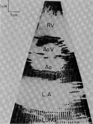
Left: Bom et.al. 1973 from ref.27. Right: Griffith and Henry 1974 from ref.30.


Left: Thurstone et.al. 1976 from ref.32. Center: Eggleton et.al. 1975 from ref.34. Right: Hisanaga et.al. 1978 from ref.37.


Left: Hisanaga et.al. (Transesophageal) 1977 from ref.36. Right: Hisanaga et.al. (Transesophageal) 1980 from ref.51.


Hisanaga et.al. (Transesophageal) Mitral stenosis 1980 from ref.50.


Left: Hisanaga et al. (Transesophageal) Image of LCA and RCA 1982 from ref.53.
Right: Souquet J (Transesophageal horizontal scan) Mitral valve prolapse 1982 from ref.56.
References
1. Edler I, Hertz CH. The use of ultrasonic reflectoscope for the continuous recording
of the movement of heart walls. Kungl. Fysiographiska Sollskapet i Lund Forhandlingar, Band 24, 1954, S 40-58 M-mode echo


Left: Inge G. Edler. Right: C. Hellumuth Hertz. From 1977 Albert Lasker Clinical Medical Research Award.

Example of the earliest M-mode echocardiogram. This image was probably recorded in 1953 or 1954.
From Roelandt JR Short history of cardiac ultrasound. Eur J Echocardiography, 1:8-11, 2000

M-mode system developed by Edler and Hertz. (Acta Med Scand Suppl. 1961;370:5-123)
2. Satomura S, Matsubara S, Yoshioka M. A new method of mechanical vibration
measurement and its application. Mem. Inst. Sci. Ind. Res. Osaka Univ. 13:125-133,1956
Continuous wave Doppler echo

Shigeo Satomura, PhD.
From Short history of the development of ultrasound by Joseph Woo.
2A. Wild JJ, Crawford HD, Reid JM. Visualization of the excised human heart by means of reflected ultrasound or echography: Preliminary report. Am Heart J 54(6):903-906, 1957
3. Edler I, Gustafson A. Ultrasonic cardiogram in mitral stenosis; Preliminary communication. Acta Med Scand 159:85-90, 1957 Continuous wave Doppler echo
4. Satomura S. Ultrasonic Doppler method for the inspection of cardiac function. J Acoust Soc Am 29:1181-1185, 1957
4A. Satomura S. Study of the flow patterns in peripheral arteries by ultrasonics.
J acoust Soc Jpn 15:151-158, 1959
5. Cieszynski T. Intracardiac method for the investigation of structure of the heart with the aid of ultrasonics. Arschiwum Immunologii i terapii Doswiadczalnej 8:551-557, 1960
Examination of animal (dog). A-mode. Catheter head ultrasound probe.
6. Olofsson S. An ultrasonic optical mirror system. Acoustica 13:361-367, 1963


(a) (b)
(a) A two-dimensional ultrasonic picture of an isolated heart of a calf.
(b) After the experiment, the heart was cut along the plane which was transversed by the ultrasound.
(a)(b) from ref.6.
7. Hertz CH, Äsberg AG. 5th International Conference on Medical Electronics in Liege, Belgium in July 1963. The first report of two-dimensional images of the human heart in vivo.

From page 1605 in Edler's paper: Edler I, Lindstrom K. The history of echocardiography.
Ultrasound in Medicine and Biology, Vol.30:12, pp1565-1644, 2004
(This is photography of Edler's paper) Please see ref.8.
Hertz reported two-dimensional echocardiography of the living human heart for the first time in history.
8. Hertz CH. Ultrasonic heart investigation. Med Electron Biol Engng Vol.2, pp39-45, Pergamon Press,1964.
This paper is based on a paper(ref.7) presented at the 5th International Conference on Medical Electronics in Liege, Belgium in July 1963. Please see ref.7.


From ref.8 Two-dimensional image of the human heart in vivo taken with the mirror system. Horizontal scan. This is the first full paper of real-time two-dimensional echocardiography of the living human heart. Original image is more beautiful than this photography.
9. Omoto R, Atsumi K, Hori M, Suma K, Toyoda T, Sakurai Y, Muroi T, Fujimori Y, Uchiyama A, Uchida R and Nagasaki K. Ultrasonic Intravenous Sonde. Japanese Journal of Medical Electronics and Biological Engineering 1, 90, 1963(In Japanese) Linear sweeping by pushing and pulling catheter.
9A. Joyner CR Jr, Reid JM, Bondb JP. Reflected ultrasound in the assessment of mitral valve disease. Circulation 27:503-511, 1963 M-mode.
10. Hertz CH, Olofsson S. A mirror system for ultrasonic visualization of soft tissues.
In Kelly E (ed.) Symposium on ultrasound in Biology and Medicine, Ultrasonic Energy, University Illinois Press 322-326, 1964
11. Äsberg AG. Ultrasonic cinematography of the living heart. Arkiv for Fysik, 30:536-537, 1965 Please see ref.7
11A. Feigenbaum H, Waldhausen JA, Hyde LP. Ultrasound diagnosis of pericardiac effusion. JAMA 191: 711-714, 1965 M-mode. Human.
12. Hertz CH. Ultrasonic engineering in heart diagnosis. Am J Cardiol 19:6-17, January 1967

C. Hellmuth Hertz. From 1977 Albert Lasker Clinical Medical Research Award.

From ref.12 Images of normal human heart. The movement of the mitral valve can be followed through the cardiac cycle.
13. Äsberg AG. Ultrasonic cinematography of the living heart. Ultrasonics 5:113, 1967
14. Omoto R. Intracardiac scanning of the heart with the aid of ultrasonic intravenous probe. Jp Heart J 1967, 8:569-581

From ref.6 Records of ECG gated linear scanning by pushing and pulling catheter very slowly.
(a) A 24-year-old male with ASD.
(b) A 20-year-old female with ASD.
(c) A 14-year-old male with ASD.
(d) A 15-year-old male who is suspected of ASD.

Ryozo Omoto, MD. From "People who developed medical ultrasound". p71, Itoh K, Ed., Supplement of Proceedings of 60th Meeting of JSUM 1992
15. Ebina T, Oka S, Tanaka M, Kosaka S, Terasawa Y, Unno K, Kikuchi Y, Uchida R. The ultrasono-tomography for the heart and great vessels in living human subjects by means of the ultrasonic reflection technique. Jpn Heart J 1967, 8:331-353
Compound electrocardiographic gated cross-sectional imaging of heart.

Toshiaki Ebina, MD From "Published to mark retirement from Tohoku University".
16. Baker DW, Watkins D. A phase coherent pulse Doppler system for cardiovascular measurement. Proc 20th Ann Conf on Eng in Med and Biol, Boston, paper 27-2 (abstr), November 1967

Donald Baker From "History of Ultrasound in Obstetrics and Gynecology".
17. Somer JC. Instantaneous and continuous pictures obtained by a new two-dimensional scan technique with a stationary transducer. Proceedings in Echo-Encephalography -International Symposium on Echo-Encephalography, (Eds.) Kazner E, Schiefer W, Zulch KJ, Publishers Springer Verlag, New York, pp234-238, 1967 Phased array sector scanner
18. Somer JC. Electronic sector scanning for ultrasonic diagnosis. Ultrasonics 6:153-159, 1968 Phased array sector scanner
18A. Light LH Non-injurious Ultrasonic Technique for observing Flow in the human Aorta. Nature 224: 1119-1121, 1969
19. Somer JC. Electronic sector scanning with ultrasonic beams. In: Ultrasonographia Medica Vol.1, Proceedings of the First World Congress on Ultrasonic Diagnostics in Medicine and SIDUO III, (Eds.) Bock J, Ossoinig K, Publ. Verlag der Wiener Medizinisehen Akademie,
Wien, pp27-32, 1971 Phased array sector scanner

Jan C Somer. From Short history of the development of ultrasound by Joseph Woo.

Somer's phased array probe . From Short History of the development of ultrasound by Dr. Joseph Woo.
20. Contrast echo was first observed by Claude Joyner and reported orally. Gramiak heard that.
21. Gramiak R, Shah PM. Echocardiography of the aortic root. Investigative Radiology 3(5):356-366, 1968
21A. Gramiak R, Shah PM and Krammer DH. Ultrasound cardiography: Contrast studies and function. Radiology 92:939-948, 1969


Left: Raymond Gramiak from Karamanou M, Papaioannou TG, Stefanadis C, Androutsos G
Genesis of ultrasonic microbubbles: a quick historical overview, Current Pharmaceutical Design, 8:2115-
2117, 2012
Right: Pravin M Shah from Cary Cardiology, Intersocietal Accreditation Commission.
22. Wells PNT. A range-gated ultrasonic Doppler system. Med Biol Eng 7:641-652, 1969
23. Baker DW. Pulsed ultrasonic Doppler blood-flow sensing. IEEE Transactions on Sonics and Ultrasonics Vol.17 Issue:3 pp170-184, 1970 See ref.16.
Pulsed Doppler was reported by DW Baker 2 years before report of Wells (ref.16).
24. Side CD, Gosling RG. Nonsurgical assessment of cardiac function. Nature 232: 335-336, 1971 Esophageal continuous wave Doppler echo

From ref.24 Intraesophageal method. Continuous wave Doppler only. Special analysis of Doppler shift signals from descending thoracic Aorta. Comment: The inspection target was not written in this paper. We could not know whether this image was a human's or an animal's.
25. Bom N, Lancee CT, Honkoop J, Hugosholy PG. Ultrasonic viewer for cross-sectional analyses of moving cardiac structures. Biomedical Eng. 6:500-508, 1971 Linear scanner.
26. King DL. Cardiac ultrasonography. A stop-action technique for imaging intracardiac anatomy. Radiology 103(2):387-392 ,1972 Compound electrocardiographic gated cross-sectional image of heart
26A. Johnson ML, Holmes JH, Spangler RD, Paton BC Usefulness of echocardiography in patients undergoing mitral valve surgery. J Thorac Cardiovasc Surg 64(6): 922-934, 1972
27. Bom N, Lancee CT, van Zwieten G, Kloster FE, Roelandt J. Multiscan echocardiography I, Circulation 48:1066-1074, 1973 Linear scanner.

Nicolaas Bom. From "Technology of Real-Time Ultrasound" by N. Bom.
From ref.27 Bom's Linear array transducer.

From Multiscan Echocardiography II. Technique and Initial Clinical Results. Circulation 48:1075-1084, 1973
Sagital cardiac cross-section obtained with oblique transducer position.
27A. King DL. Real-time cross-sectional ultrasonic imaging of the heart using linear array multi-element transducer. J Clin Ultrasound 1(3):196-201, 1973
28. Griffith JM and Henry WL. A real time system for two-dimensional echocardiography. Proceedings of 20th Annual Conference on Engineering in Medicine and Biology, Minneapolis, 15:423, 1973 Mechanical sector scanner

Walter L Henry M.D. From History of Echocardiography, NEW AND AMAZING dot COM.
28A. Griffith JM, Henry WL, Epstein SE. Real time two-dimensional echocardiography. Circulation 48(Suppl. Ⅳ):124, 1973
28B. Johnson SL, Baker DW, Lute RA, Dodge HT. Doppler echocardiography: The localization of cardiac murmurs. Circulation 48(4):810-822, 1973
29. Gramiak R, Waag R, Simon W. Cine ultrasound cardiography. Radiology, 107:175-180, 1973

From ref.29 Pseud 2D image using a spatially oriented reconstruction of M-mode echogram.
30. Griffith JM, Henry WL. A sector scanner for real time two dimensional echocardiography. Circulation 49:1147-1152, 1974

From ref.30 Mechanical sector scanner.

From ref.30 Horizontal scan at the level of aortic valve.
31. von Ramm OT, Thustone FL, Kisslo J. Real time two-dimensional echocardiography.
Spie Seminar Proceedings 47:93-95, 1974 Phased array sector scanner

Olaf T. von Ramm from Duke University, Pratt school of Engineering.
32. Thurstone FL, von Ramm OT. A new ultrasound imaging technique employing two- dimensional electronic beam steering. In Green PS (ed.) Acoustical Holography, New York, Plenum Press pp149-159, 1974 Phased array sector scanner

Fredrick L Thurstone from the Orlando Sentinel on Mar. 24, 2005

From The paper: Cardiac Imaging Using a Phased Array Ultrasound System II. Clinical Technique and Application, Circulation 53:262-267,1976 Phased array transducer and a patient.

From The paper: Cardiac Imaging Using a Phased Array Ultrasound System II. Clinical Technique and Application, Circulation 53:262-267,1976
Image is a Photo from a stop action video tape frame through the long axis of the left ventricle.
32A. Teichholz LE, Cohen MV, Sonnenblick EH, Gorlin R. Study of left ventricular geometry and function by B-scan ultrasonography in patients with and without asynergy. N Engl J Med 291(23):1220-1226, 1974
33. Eggleton RC, Johnston KW. Real time mechanical scanning system. Proceedings SPIE Annual Meeting, August 1974 Mechanical sector scanner
33A. Huntsman LL, Gams E, Johnson CC, Fairbanks E. Trancutaneous determination of aortic blood-flow velocities in man. Am Heart J 89(5):605-612, 1975
33B. Holm HH, Kristensen JK, Pedersen JF, Hancke S, Northeved A. A new mechanical real time ultrasonic contact scanner. Ultrasound in Medicine and Biology 2(1):19-23, 1975
34. Eggleton RC, Feigenbaum H, Johnston KW et. al. Visualization of cardiac dynamics with real-time B-mode ultrasonic scanner. In White D (ed.) Ultrasound in Medicine Vol. 1, Plenum Press, New York pp385-393, 1975 Mechanical sector scanner

From ref.34 The sector scanner and electronic module.

From ref.34 Long axis cross-section through aortic valve in normal individual on left and a patient with aortic stenosis on right.
34A. Nealeigh RC, Miller CW. A venous pulse Doppler catheter tip flowmeter for measuring arterial blood velocity, flow, and diameter in deep arteries. ISA trans. 15(1):84-87, Jan 1976
34B. Holen J, Aaslid R, Landmark K, Simonsen S. Determination of pressure gradient in mitral stenosis with a non-invasive ultrasound Doppler technique. Acta Med Scand 119(6):455-460, 1976
34C. Weyman AE, Feigenbaum H, Dillon JC, Johnston KW, Eggleton RC. Noninvasive visualization of the left main coronary artery by cross-sectional echocardiography. Circulation 54(2):169-174, 1976
35. Frazin L, Tarano JV, Stephanides L, Loeb HS, Kopel L, Gurnar RM. Esophageal echocardiography. Circulation 54:102-108, 1976 Esophageal M-mode echocardiography

From ref.35 Photograph of esophageal transducer. M mode only.

From ref.35 Panel A shows the external echo of a patient with documented mitral stenosis. Panel B shows the esophageal echo counterpart with reversed orientation.
35A. Hisanaga K, Hisanaga A, Nagata K, Yoshida S. Improvement of ultra-wide angle sector scanning system and its clinical application. Proceedings of the Japan Society of Ultraonics in Medicine 32:47-48, 1977

From ref.35A Entire adult heart image.
36. Hisanaga K, Hisanaga A, Nagata K, Yoshida S. A new transesophageal real time two-dimensional echocardiographic system using a flexible tube and its clinical application.
Proceedings of the Japan Society of Ultraonics in Medicine 32:43-44, 1977
This is the first transesophageal 2D scanner in history. The Term “Transesophageal” was first used in this paper. On the other hand, Frazin used Term “Esophageal”.

From ref.36 Transesophageal horizontal scan in a 26 years old female.

Kohzoh Hisanaga, MD. (Hisanaga K.) From "People who developed medical ultrasound". p35, Itoh K, Ed., Supplement of Proceedings of 60th Meeting of JSUM 1992. Kohzoh Hisanaga is not only an electronic engineer but also a medical doctor.
Dr. Hisanaga received the Honor Award of the Japanese college of cardiology in 1991 because Dr. Hisanaga developed Transesophageal two-dimensional echocardiography for the first time in history.
36A. Hatle L, Brubakk A, Tromsdal A, Angelsen B. Noninvasive assessment of pressure drop in mitral stenosis by Doppler ultrasound. Br Heart J 40(2):131-140. 1978
37. Hisanaga K, Hisanaga A. A new real-time sector scanning system of ultra-wide angle and real-time recording of entire adult cardiac images --Transesophagus and Trans- chest- wall methods --. In White DN, Lyons AE (eds.) Ultrasound in Medicine Vol.4, New York, Plenum Press 1978, pp391-402

From ref.37 Insertions of transducer to esophagus and transesophageal ultrasound examinations were performed with patients in left lateral position(left). Typical horizontal scan in a 26-year-old normal adult by using transesophageal method(right).

From ref.37 Transthoracic image. A long axis scan in a 31-year-old normal man. Entire heart image is seen. The endocardium of the left ventricle and the right ventricular anterior wall are seen.

From ref.37 Transesophageal M-mode echograms. These images were recorded in order to identify echo sources of transesophageal cross-sectional images.
38. Hisanaga K and Hisanaga A. A transesophageal real-time sector scanner with an oil filled cell. Proceedings of the 23rd Annual meeting of American institute of Ultrasound in Medicine, p47, San Diego, 1978


From ref.38. Upper: Diagramattic illustration of the scanner. Lower: Horizontal scan in a normal adult subject.
39. Hisanaga K, Hisanaga A, Nagata K, Ichie Y, Yoshida S. A new transesophageal high speed linear scanner and its clinical application. Proceedings of the Japan Society of Ultrasonics in Medicine 33:47-48, 1978

From ref.39 Transesophageal vertical scan through mitral valve in a normal adult.
40. Brandestini M. Topoflow - A digital full range Doppler velocity meter. IEEE Transactions on Sonics and Ultrasonics, Vol.25(5):287-292, 1978
M-mode and multigated Doppler flowmeter.
41. Hisanaga K, Hisanaga A, Nagata K, Ichie Y, Hibi N, Fukui Y, Nishimura K, Kambe T.
A transesophageal Pulsed Doppler Echocardiographic system and initial clinical results.
Proceedings of the Japan Society of Ultrasonics in Medicine 34:9-10, 1978
Transesophageal pulsed Doppler with M-mode. This was the first human transesophageal pulsed Doppler examination in history.
41A. Griffith JM, Henry WL. An ultrasound system for combined cardiac imaging and Doppler blood flow measurement in man. Circulation 57:925-930, 1978
41B. Barash PG, Glanz S Katz JD, Taunt K, Talner NS. Ventricular function in children during halothane anesthesia: an echocardiographic evaluation. Anesthesiology 49:79-85, 1978
42. Hisanaga K, Hisanaga A, Ichie Y, Hibi N, Nishimura K, Fukui Y, Kambe T.
Transesophageal pulsed Doppler echocardiography. Lancet 1:53-54, 1979
Transesophageal pulsed Doppler with M-mode.
43. Hisanaga K and Hisanaga A. A new transesophageal radial scanner using a rotating flexible shaft and initial clinical results. Proceedings of the 24th Annual meeting of American institute of Ultrasound in Medicine, p122, Montreal, August, 1979


From ref.43 left : Diagram of transesophageal radial scanner.
right : A horizontal scan in a normal adult woman by using the transesophageal radial scanner.
44. Hisanaga K, Hisanaga A, Nagata K, Ichie Y. Cardiac imaging using a transesophageal ultrasound high speed rotating scanner. Proceedings of the Japan Society of Ultrasonics in Medicine 35:157-158, 1979
Photography of the original paper


From ref.44 Transesophageal high speed rotating scanner.

From ref.44 Transducer and commutator in oil bag.

From ref.44 Horizontal scan at the level of the mitral valve in a patient with severe mitral stenosis by using the transesophageal high speed rotating scanner. Anterior and posterior mitral leaflets are thickened.
45. Hisanaga K, Hisanaga A, Ichie Y. A transesophageal ultrasound sector scanner for oblique scan. (abstr) Circulation 60 (Suppl.Ⅱ): Ⅱ-245.1979
46. Holen J, Simonsen S. Determination of pressure gradient in mitral stenosis with Doppler echocardiography. Br Heart J 41:529-535, 1979
46A. Hatle L, Angelsen BA, Tromsdal A. Non-invasive assessment of aortic stenosis by Doppler ultrasound. Br Heart J 43(3):284-292, 1980
47. Hisanaga K, Hisanaga A, Kambe T. Detection of atrial septal defect by transesophageal two-dimensional echocardiography(abstr). Circulation 62(supple Ⅲ): Ⅲ-34, 1980
48. Hisanaga K, Hisanaga A, Nagata K, Ichie Y. A transesophageal high speed rotating scanner for oblique scan and long axis cardiac images including apex. Proceedings of the Japan Society of Ultrasonics in Medicine 36:395-396, 1980
Four chamber view including apex could be observed easily by this system.

From ref.48 Inferior oblique scan through the apex in a normal adult. Cross-section is angled downward about 40 degrees from the horizontal plane. The entire heart including the apex is seen.
49. Brandestini MA, Eyer MK, Stevenson JG. M/Q mode echocardiography: the synthesis of conventional echo with digital multigated Doppler. In Echocardiography, edited by Lancee CT. Martinus Nijhoff Publishers, The Hague, pp441-446, 1979
M-mode and multigated Doppler flowmeter.
50. Hisanaga K, Hisanaga A, IchieY, Hibi N, Nishimura K, Kambe T. High speed rotating
scanner for transesophageal cross-sectional echocardiography. Am J Cardiol 46:837-842, 1980

From ref.50 Diagrammatic illustration of the transesophageal high speed rotating scanner. A small transducer in the esophagus is rotated through a full 360° through a flexible shaft by a motor at 15 to 50 cycles/s. Although the small transducer is rotated with great speed in the patient's esophagus, no damage results because the transducer is safely enveloped in an oil bag.

From ref.50 Transesophageal high speed rotating scanner.

From ref.50 Transducer and commutator in oil bag. Sound energy is coupled to and from the transducer through the slip-ring commutator because of the full 360°rotation of the transducer.


From ref.50 Transesophageal cross-sectional echocardiograms in a patient with mitral stenosis. The cross section is horizontal and shows the heart as viewed from the cardiac apex. A: a frame during diastole and B: a frame during systole. A stenotic mitral orifice (in A) is seen between the tips of the thickened mitral leaflets. The interatrial septum (IAS) is seen without dropout. AML= anterior mitral leaflet, ESO = esophagus, IVS = interventricular septum, LA = left atrium, LV = left ventricle, PML = posterior mitral leaflet, RA = right atrium, RV = right ventricle, TV = tricuspid valve.
51. Hisanaga K, Hisanaga A, Nagata K, IchieY. Transesophageal cross-sectional echocardiography. Am Heart J 1980, 100:605-609

From ref.51 Transesophageal horizontal scan at the level of the aortic valve in a patient with mitral stenosis. The aortic cusps are closed in diastole. Large left atrium is seen. Right ventricular outflow tract is seen anterior to the aorta. AV = aortic valve, RVOT = right ventricular outflow tract.

From ref.51 Transesophageal vertical linear scan through the pulmonary artery in a normal subject. Bifurcation of pulmonary artery and part of ascending aorta are seen. AO = aorta, PA = pulmonary artery, BI = bifurcation of pulmonary artery.
51A. Spotnitz HM, Young CYH et al. Intraoperative left ventricular performance evaluated by two-dimensional ultrasound. Circulation 62:329, 1980
52. Hisanaga K, Hisanaga A, Isaji F. Transesophageal two-dimensional echocardiographic diagnosis of left atrial myxoma. Proceedings of the Japan Society of Ultrasonics in Medicine 39:457-458, 1981

From ref.52 Transesophageal two-dimensional echocardiogram in a patient with a left atrial myxoma. Cross section is horizontal. A stalk of the tumor is seen clearly. K = stalk.

From ref.52 Extracted tumor. Weight of the tumor was 38.5g.
52A. Sahn DJ. Intraoperative Applications of Two-Dimensional and Contrast Two-Dimensional Echocardiography for Evaluation of Congenital, Acquired and Coronary Heart Disease in Open-Chested Humans during Cardiac Surgery. In Echocardiography, edited by Rijsterborgh H. The Hague, Martinus Nijhoff Publishers pp9-23, 1981
52B. Sponitz HM. Two-dimensional ultrasound and cardiac operations. J Thoracic Cardiovasc Surg 83(1):43-51, 1982
53. Hisanaga K, Hisanaga A. Findings of coronary artery by transesophageal two-dimensional echocardiography. Proceedings of the Japan Society of Ultrasonics in Medicine 40:171-172, 1982

From ref.53 Transesophageal two-dimensional echocardiogram in a patient with both aortic regurgitation and mitral stenosis. LCA = left coronary artery, RCA = right coronary artery.
54. Ghosh A, Nanda NC, Maurer G. Three-dimensional reconstruction of echocardiographic images using the rotational method. Ultrasound in medicine and biology 8(6):655-661, 1982
55. Schiller NB. Evaluation of cardiac function during surgery by transesophageal 2-dimensional echocardiography. In: Hanrath P, Bleifeld W, Souquet J (eds.) Cardiovascular Diagnosis by Ultrasound, Martinus Nijhoff Publishers pp289-293, 1982
55A. Seward JB, Tajik AJ, Dimagno EP. Esophageal phased-array sector echocardiography. In: Hanrath P, Bleifeld W, Souquet J (eds.) Cardiovascular Diagnosis by Ultrasound, Martinus Nijhoff Publishers pp270-279, 1982 Not human but animal.
55B. Souquet J, Hanrath P, Ziteli L, Kremer P, Langenstein BA, Schluter M.
Transesophageal phased array for imaging the heart. IEEE Transactions on Biomedical Engineering 29:707-712, 1982

Jacques Souquet, PhD. From Eugene A Hessel et. al. Evolution of Perioperative Echocardiography. Anesthesia Key.
56. Souquet J. Phased array transducer technology for transesophageal imaging of the heart -current status and future aspects-, In Hanrath P, Bleifeld W and Souquet J(eds.), Cardiovascular Diagnosis by Ultrasound, Martinus Nijhoff Publishers pp251-259, May, 1982.
Transesophageal phased array sector scanner.

From ref.56 Mitral valve prolapse.
56A. Hanrath P, Schluter M, Langenstein BA, Polster J, Engel S. Transesophageal horizontal and sagittal imaging of the heart with phased array system. Initial clinical results. In Hanrath P, Bleifeld W and Souquet J (eds.) Cardiovascular Diagnosis by ultrasound, Martinus Nijhoff Publishers 1982, pp280-288
56B. Schluter M, Langenstein BA, Polster J, Kremer P, Souquet J, Engel S, Hanrath P.
Transesophageal cross-sectional echocardiography with a phased array transducer system.
Technique and initial clinical results. Br. Heart J. 48:67-72, 1982
56C. Cahalan MK et al. Intraoperative monitoring with two-dimensional echocardiography.
Anesthesiology 57: A153, 1982 Transesophageal phased array 2D sector scanner.
Intraoperative monitoring with transesophageal 2D echo.
56D. Kremer P et al. Intraoperative monitoring of left ventricular performance by transesophageal M-mode and 2-D echocardiography. (abstr) Am J Cardiol 49:956, 1982.
Transesophageal phased array 2D sector scanner.
56E. Langenstein BA, Hanrath P, Polster J, Kremer P, Engel S, Souquet J. Detection of atrial septal defect by transesophageal cross-sectional contrast echocardiography. (abstr) Am J Cardiol 49(4):956, March 1982
57. Hisanaga K, Hisanaga A. Transesophageal cross-sectional echocardiography with a mechanical scanning system. In: Hanrath P, Bleifeld W, Souquet J (eds.) Cardiovascular Diagnosis by Ultrasound, Martinus Nijhoff Publishers pp239-246, 1982
58. Bommer WJ, Miller L. Real-time two-dimensional color flow Doppler -- Enhanced Doppler flow imaging in the diagnosis of cardiovascular disease. Am J Cardiology (Abstr) 49:944, 1982
59. Namekawa K, Kasai C, Tsukamoto M, Koyano A. Imaging of blood flow using autocorrelation. Ultrasound in Medicine and Biology 8(Suppl. Ⅰ):138,1982
Color Doppler by using a mechanical ultrasound sector scanner.
60. Namekawa K, Kasai C, Tsukamoto M, Koyano A. Real time blood flow imaging system utilizing auto-correlation techniques. In Lerski RA, Morley P (eds.) Ultrasound, New York, Pergamon Press pp203-208, 1982 Color Doppler by using a mechanical ultrasound sector scanner.

Kouroku Namekawa. From "Meet The History" Japan Heart Foundation.
Mr. Namekawa received the Honor Award of the Japanese college of cardiology in 1990.
60A. York PG, Popp RL. Noninvasive estimation of right ventricular systolic pressure by Doppler ultrasound in patients with tricuspid regurgitation. Circulation Oct 70(4) 657-662, 1984
61. Kasai C, Namekawa K, Koyano A, Omoto R. Real time two-dimensional blood flow imaging using an autocorrelation technique. IEEE Transactions on Sonics and Ultrasonics 32(3):458-464, May 1985 Color Doppler by using a phased array ultrasound sector scanner.

Chihiro Kasai. From Kobayasi Riken News No.67-6
61A. Trahey GE, Smith SW. Properties of acoustical speckle in the presence of phase aberration. Part I: First order statistics. Ultrasonic Imaging 10:12-28,1988
61AA. Pandian NG, Krevis A, Brockway B, Isner JM, Sacharoff A, Boleza E, Caro R, Muller D. Ultrasound angioscopy: real-time two-dimensional intraluminal ultrasound imaging of blood vessels. Am J Cardiol 62:493-494, 1988
61B. Smith SW, Trahey GE, Hubbard SM, Wagner RF. Properties of acoustical speckle in the presence of phase aberration. Part II: Correlation lengths. Ultrasonic Imaging 10:29-51, 1988
61C. Bertrand M, Meunier J, Doucet M, Ferland G. Ultrasonic biomechanical strain gauge based on speckle tracking. Proceedings, 1989 IEEE Ultrasonic Symposium, pp859-864, 1989
61D. Meunier J, Bertrand M. Ultrasonic speckle motion inherent to tissue motion: theory and simulation. Proceedings, 1989 IEEE Ultrasonic Symposium, pp865-868, 1989
62. Wollschlager H, Zeiher AM, Klein HP, Kasper W, Wollschlager S, Geibel A, Just H.
"Transesophageal echo computer tomography (ECHO-CT): a new method of dynamic 3-D reconstruction of the heart)" (in German) Biomed. Tech. (Berl) 34 Suppl:10-1, 1989
63. Nissen SE, Gurley JC, Grines CL, Booth DC, McClure R, Berk M, Fischer C, DeMaria AN. Intravascular ultrasound assessment of lumen size and wall morphology in normal subjects and patients with coronary artery disease. Circulation 1991, 84:1087-1099
64. von Ramm OT, Smith SW, Pavy HG Jr. High speed ultrasound volumetric imaging system. Part II: Parallel processing and image display. IEEE Transactions on Ultrasonics Ferroelectrics, and Frequency Control 38:109-115, 1991
64A. McDicken WN, Sutherland GT et al. Color Doppler velocity imaging of the myocardium. Ultrasound Med Biol 18: 651-654, 1992 Color Doppler myocardial imaging.
Tissue Doppler Imaging.
64AA. Wells PNT. Milestones in cardiac ultrasound: echoes from the past. History of cardiac ultrasound. Int J Card Imaging 9 (Suppl 2):3-9, 1993
64B. Tei C. New non-invasive index for combined systolic and diastolic ventricular function. J Cardiol 26(2):135-6, 1995
64C. Tei C, Ling LH, Hodge DO, Bailey KR, Oh JK, Rodeheffer RJ, Tajik AJ, Seward JB. New index of combined systolic and diastolic myocardial performance: a simple and reproducible measure of cardiac function--a study in normals and dilated cardiomyopathy. J Cardiol 26(6):357-366, 1995
65. Roelandt J, ten Cate FJ, Bruining N, Salustri A, Vietter WB, Mumm B, van der Putten N.
Transesophageal rotoplane echo-CT. A novel approach to dynamic three-dimensional echocardiography. Thoraxcentre J 6:4-8, 1993

Jos Roelandt from "EuroEcho Budapest, Hungary Day 3 by James Thomas."
65A. Sutherland GR, Stewart MJ et al. Color Doppler myocardial imaging: a new technique for the assessment of myocardial function. J Am Soc Echocardiogr 7(5): 441-458, 1994 Color Doppler myocardial imaging.
65B. Paika P, Lange A et al. Doppler tissue imaging: Myocardial wall motion velocities in normal subjects. J Am Soc Echocardiogr 8: 659-668, 1995
65C. Miyatake K, Yamaguchi M et al. New method for evaluation left ventricular wall motion by color-coded tissue Doppler imaging: In vitro and in vivo studies. J Am Coll Cardiol 25(3): 717-724, 1995
65D. Sugeng L, Cao QL et al. Three-dimensional echocardiographic evaluation of aortic disorders with rotational multiplanar imaging; experimental and clinical studies. J Am Soc Echocardiogr 10(2): 120-132, 1997
65E. Heimdal A, Stoylen A, Torp H, Skjaerpe T. Real-time strain rate imaging of the left ventricle by ultrasound. J Am Soc Echocardiogr 11:1013-1019, 1998
66. Kisslo J, Firek B, Ota T, Kang DH, Fleishman CE, Stetten G, Li J, Ohazama CJ, Adams D, Landlfo C, Ryan T, von Ramm OT. Real time volumetric echocardiography. The technique and possibilities. Echocardiography 17:773-779, 2000 Real-time 3D echo. Matrix array transducer.
67. Ota T, Kisslo J, von Ramm OT, Yoshikawa J. Real-time, volumetric echocardiography: usefulness of volumetric scanning for the assessment of cardiac volume and function. J Cardiol 37 Suppl 1:93-101, 2001 Real-time 3D echo. Matrix array transducer.
67A. Weidemann F, Eyskens B, Mertens L, Dommke C, Kowalski M, Simmons L, et Claus P, Bijnens B, Gewillig M, Hatle L, Sutherland GR. Quantification of regional right and left ventricular function by ultrasonic strain rate and strain indexes after surgical repair of tetralogy of Fallot. Am J Cardiol 90(2):133-138, 2002
68. Franke A, Kuhl HP. Second-generation real-time 3D echocardiography a revolutionary new technology. MEDICA MUNDI 47/2, August 2003. Second generation real-time 3D echo. Matrix array transducer.

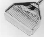
From ref.68 Fig.1 Microscopic view of a matrix array transducer. The size of a human hair is shown for comparison (arrows).
68A. Leitman M, Lysyansky P, Sidenko S, Shir V, Peleg E, Binenbaum M, Kaluski E, Krakover R, Vered Z. Two-dimensional strain-a novel software for real-time quantitative echocardiographic assessment of myocardial function. J Am Soc Echocardiogr 17(10): 1021-1029, 2004
69. Edler I, Lindstrom K. The history of echocardiography. Ultrasound Med Biol 2004, 30:1565-644
70. Ohtsuki S, Tanaka M. The flow velocity distribution from Doppler information on a plane in three-dimensional flow. Journal of Visualization 9(1):69-82, 2006
71. Salgo IS. 3D echocardiographic visualization for intracardiac beating heart surgery and intervention. Seminars in Thoracic Cardiovascular Surgery 19:325-329, 2007
Transesophageal 3D echo. Matrix array probe.
72. Sugeng L, Sheman SK, Salgo IS, Weinert L, Shook D, Ramen J, Jeevanandam V, Dupont F, Settlemier S, Savord B, Fox J, Mor-Avi V, Lang RM. Live 3-dimensional transesophageal echocardiography initial experience using fully-sampled matrix array probe. Journal of the American College of Cardiology 52:446-449, 2008 Transesophageal 3D echo. Matrix array probe.
73. Garcia D et al. Two-dimensional intraventricular flow mapping by digital processing conventional color-Doppler echocardiography images. IEEE Trans Med imaging 29:1701-
1713, 2010 VFM. Vector Flow Mapping.
74. Voigt JU, Pedrizzetti G, Lysyansky P et al. Definitions for a common standard for 2D speckle tracking echocardiography: consensus document of the EACVI/ASE/Industry Task Force to standardize deformation imaging. Eur Heart J Cardiovasc Imaging 16(1):1-11, January 2015 Speckle tracking.
75. Dietrich CF, Bolondi L, Duck F et al. History of ultrasound in Medicine from its birth to date(2022), on occasion of the 50 Years Anniversary of EFSUMB. Med Ultrason 0:1-19, 2022 Online first
76. Fraser AG, Monaghan MJ, van der Steen AFW, Sutherland GR. A concise history of echocardiography: timeline, pioneers, and landmark publications. European Heart Journal - Cardiovascular Imaging 00:1–14, 2022 The most detailed history of echocardiography.

From ref.76 Page 1. Graphical abstract. Hisanaga’s research performance of transesophageal imaging is reported in this abstract.