History of filled water method in endoscopic ultrasonography.
History of water filled stomach method in endoscopic ultrasonography.
History of filling stomach with water method in endoscopic ultrasonography.
History of water filled balloon around the transducer method in endoscopic ultrasonography.
In endoscopic ultrasound examinations, when we put the transducer on the stomach wall directly, it is sometimes difficult for us to obtain images of diagnostic quality. In order to increase image quality of EUS, we must use water filled stomach method or water filled balloon around the transducer method. These are the most useful discoveries in the history of EUS examination.
Hisanaga developed and used both water filled stomach method(ref.12) and water filled balloon (oil filled bag) method (ref.3A, ref.5, ref.6, ref.9, ref.10) for the first time in the world.
By using these methods we can obtain high quality images of abdomen. Nowadays these methods are indispensable in the EUS examinations.
I and my coworker medical doctors performed this methods by themselves and I knew this methods were not dangerous. However when I reported water filled stomach method for the first time in the world in 1980, famous Japanese professor objected to this method because water filled stomach methods must be very dangerous for patients. I was very surprised when I knew that 4 years after my report, this professor reported water filled stomach method by himself in 1984.
If you have a time please see
"History of endoscoic ultrasonography" Click here
"History of endoscopic ultrasonography (EUS) and Transesophageal echocardiography (TEE)" Click here
"History of echocardiography" Click here
References
1. Hisanaga K, Hisanaga A, Nagata K, Yoshida S A new transesophageal real time two-dimensional echocardiographic system using a flexible tube and its clinical applications. Proceedings of Japan Society of Ultraonic in Medicine 32:43-44, 1977

From Ref.1 Transesophageal horizontal scan in a normal adult.
2. Hisanaga K, Hisanaga A. A new trans-digestive-tract scanner with a gastrofiberscope. Proceedings of the 23rd Annual Meeting of American Institute of Ultrasound in Medicine. p.108, November, San Diego, 1978
3. Hisanaga K, Hisanaga A. A new real-time sector scanning system of ultra-wide angle and real-time recording of entire adult cardiac images --Transesophagus and Trans-chest-wall methods --. In:White DN, Lyons AE, eds. Ultrasound in Medicine. Vol.4. New York; Plenum Press, pp391-402, 1978
From Ref.3 Insertions of transducer to esophagus and transesophageal ultrasound examinations were performed with patients in left lateral position(left). Typical horizontal scan in a normal adult by using transesophageal method(right).

From Ref.3 A long axis scan in a 31-year-old normal man. Entire heart image is seen. The endocardium of the left ventricle and the right ventricular anterior wall are seen.

From Ref.3 Transesophageal M-mode echograms. These images were recorded in order to identify echo sources of transesophageal cross-sectional images shown in Fig.10. Arrows A,B and C of Fig.10A and Fig.10D correspond to Fig.9A, 9B and 9C in each and show directions of M-mode echograms.
3A. Hisanaga K, Hisanaga A transesophageal real-time sector scanner with an oil filled cell. Proceedings of the 23rd Annual Meeting of American Institute of Ultrasound in Medicine. p.47, November, San Diego, 1978
4. Hisanaga K, Hisanaga A, Nagata K, Ichie Y. A trans- stomach wall sector scanner with a gastrofiberscope. Abstract of 2nd WFUMB, p383, July 22-27, Miyazaki, 1979
5. Hisanaga K, Hisanaga A, Nagata K, Ichie Y. A trans- stomach wall high speed rotating scanner and initial clinical results. Proceedings of the Japan Society of Ultrasoics in Medicine 35:115-116,1979

Photography of original paper.

From Ref.5 Trans-stomach-wall high speed rotating scanner.

From Ref.5 Horizontal scan through the left kidney in a normal adult by using the trans-stomach-wall rotating scanner. When
near gain is standard, pancreas is seen as echo free space near the stomach.
6. Hisanaga K, Hisanaga A. High speed rotating scanners for trasesophageal echocardiography and transgastoric sonography. Eizou Jouhou Medical 11:1094-1099, 1979 (In Japanese)

From Ref.6 Transgastric high speed rotating scanner with flexible tube.

From Ref.6 Horizontal scan at the level of kidney in a normal adult. Pancreas is seen very clearly.
7. Hisanaga K, Hisanaga A. Pancreatic echography using a trans-stomach wall ultrasound rotating scanner (abstr). Gastroenterology 78:1183, 1980
8. Hisanaga K, Hisanaga A, Kambe T. An endoscopic ultrasound scanner for abdominal echography (abstr). Gastrointestinal Endoscopy 26:68, 1980
9. Hisanaga K, Hisanaga A, Nagata K, Ichie Y. High speed rotating scanner for transgastoric sonography. Am.J.Roentgenol. 135:625-629, 1980

From Ref.9 Upper Fig. Intragastric high speed rotating scanner. Small transducer in stomach is rotated by flexible rotating shaft and motor at 15-50 cycles/sec.Lower Fig. Transducer and commutator in oil bag.


From Ref.9 Horizontal scans through left kidney in normal adult with intragastric high speed rotating scanner. Left : Left kidney and abdominal aorta are seen clearly. If amplitude of near field is relatively low, pancreas is seen as anechoic space near stomach wall. Right : With increasing amplitude of near field, pancreas assumes cloudlike shape.
10. Hisanaga K, Hisanaga A, IchieY, Hibi N, Nishimura K, Kambe T. High speed rotating scanner for transesophageal cross-sectional echocardiography. Am. J. Cadiol. 46:837-842, 1980

From Ref.10 Diagrammatic illustration of the transesophageal high speed rotating scanner. A small transducerin the esophagus is rotated througha a full 360° through a flexible shaft by a motor at 15 to 50 cycles /s. Although the small transducer is rotated with great speed in the patient's esophagus, no damage results because the transducer is safely enveloped in an oil bag.

From Ref.10 Transesophageal high speed rotating scanner.

From Ref.10 Transducer and commutator in oil bag. Sound energy is coupled to and from the transducer through the slip-ring commutator because of the full 360° rotation of the transducer.


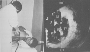
From Ref.10 Transesophageal cross-sectional echocardiograms in a patient with mitral stenosis. The cross section is horizontal and shows the heart as viewed from the cardiac apex. A : a frame during diastole and B : a frame interatrial septum (IAS) is seen without dropout. AML= anterior mitral leaflet, ESO = esophagus, IVS = interventricular septum, LA = left atrium,
LV = left ventricle, PML = posterior mitral leaflet, RA = right atrium, RV = right ventricle, TV = tricuspid valve.
11. Hisanaga K, Hisanaga A, Nagata K, IchieY. Transesophageal cross-sectional echocardiography. Am. Heart J. 100:605-609, 1980

From Ref.11 Transesphageal horizontal scan at the level of the aortic valve . The aortic cusps are closed in diastole. Large left atrium is seen. Right ventricular outflow tract is seen anterior to the aorta. AV = aortic valve, RVOT = right ventricular outflow tract.

From Ref.11 Transesophageal vertical linear scan through the pulmonary artery in a normal subject. Bifurcation of pulmonary artery and part of ascending aorta are seen. AO = aorta, PA = pulmonary artery , BI = bifurcation of pulmonary artery.
12. Hisanaga K, Hisanaga A, Kambe T. Transgastric sonography and examination technique. Proceedings of the Japan Society of Ultrasonics in Medicine 37:413-414, 1980

Photography of original paper.

From Ref.12 Horizontal scan through the stomach posterior wall in a normal adult by using the transgastric sector scanner with gastrofiberscope when the stomach was filled with water. Left kidney is seen clearly. SW = stomach wall, LK = left kidney, V = vertebra, R = right, L = left.
Content of this paper is filling stomach with water during EUS examination in order to increase acoustic contact between stomach wall and transducer. Hisanaga performed this method for the first time in the world certainly. This is the most important discovery in EUS examination.